redox

The redox landscape of SARS-CoV-2 infection
Submitted by redoxoma on
Sars-Cov-2

Main Article by Flávia Meotti (Instituto de Química da USP) and Francisco R. M. Laurindo (Incor, Faculdade de Medicina da USP)

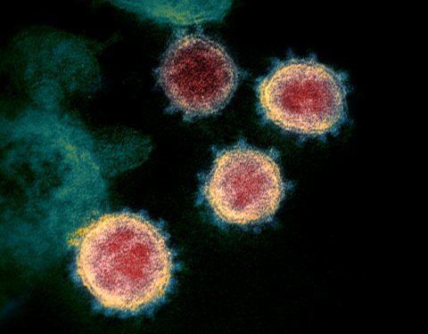
Post image credits: NIAID / CC BY

The challenge imposed by Covid-19 pandemics has promoted the union of scientists from different fields and technologies, breaking geographic frontiers to solve relevant problems. The focus is, of course, understanding SARS-CoV-2 pathobiology and the ensuing development of improved diagnostic procedures, vaccines and therapeutic compounds. Redox processes certainly belong to this story. In fact, redox pathways can be identified at essentially every level of SARS-CoV-2-related disease and hold a unique potential to integrate knowledge at different systems levels and connect multiple disciplines. Here we briefly highlight, on a somewhat arbitrary and incomplete fashion, redox phenomena of potential significance to this scenario. We hope to inspire our readers to complete this picture and pursue them in depth.

Viral entry into cells: redox and non-redox pathways

The entry of enveloped viruses such as SARS-CoV-2 into cells is a two-step process involving: (1) viral particle binding to cell-surface receptors and (2) fusion of the virion to host cell membranes.

SARS-CoV-2 binding and subsequent entry into the host cell depends essentially on the viral Spike glycoprotein (S glycoprotein). The S glycoprotein forms the characteristic corona of large distinctive spikes in the viral envelope [Xiao et al., 2003]. S glycoprotein contains the S1 domain, a globular region of the protein distal to the virus membrane and the S2 domain, which forms the stalk. S1 domain mediates high-affinity binding with the major (though not exclusive) primary cell receptor, angiotensin-converting enzyme 2, ACE2. Both glycoprotein S and ACE2 exhibit reactive cysteine residues. Membrane fusion is the result of conformational changes in virion envelope depending on its interaction with cells. For SARS-CoV-2, docking to the ACE2 induces conformational changes required for membrane fusion. For enveloped viruses including Newcastle Virus, HIV and many others, thiol/disulfide rearrangements, commented below, are essential for fusogenic activity [Fenouillet et al., 2007; Jain et al., 2008]. For the viral glycoproteins from these viruses, fusion-associated conformational changes are followed and/or preceded by disulfide bond exchanges. This could also be expected for S glycoproteins from both SARS-CoV and SARS-CoV-2, since they contain 39 Cys residues. Following this hypothesis, a series of point mutations replacing cysteines for alanines in S glycoproteins from SARS-CoV revealed that five of seven Cys present at the S1 receptor binding site are essential for association to ACE2 [Wong et al., 2004]. However, the binding of S glycoprotein to ACE2 and infection of mammal cells were largely insensitive to reducing, oxidizing or alkylating agents [Fenouillet et al., 2007; Lavillette et al., 2006]. Rather, for influenza viruses, fusion events have been linked to other stressful events such as cell acidification. These data led to the conclusion that both binding to ACE2 and the fusion capacity of the spike complex are independent of redox switches, in singular contrast to the situation described for a number of other enveloped viruses. However, recent computational simulations indicate that the affinity of SARS-CoV-2 spike protein for ACE2 is strongly impaired when the cysteines present in both proteins (not in only one of them) are in the reduced state, probably due to conformational changes, mainly in ACE2, that affect binding interaction. In contrast, binding is less affected under more oxidizing conditions able to preserve disulfides [Hati & Bhattacharyya, 2020].

Despite being poorly modulated by redox switches, cysteines present at the C-terminal region of the S2 domain undergo post-translational modifications that are crucial to virus infection. Nine of the thirty-nine cysteines in S glycoprotein are clustered between the membrane spanning domain and the C-terminal cytoplasmic tail, with six of these residues being well conserved among different coronaviruses. In SARS-CoV S glycoprotein, the cysteines clustered near to the predicted transmembrane domain were palmitoylated. Mutations of different groups of cysteines significantly decreased protein palmitoylation and virus fusion into mammal cells [Petit et al., 2007]. Since SARS-CoV-2 S glycoprotein shares 79.6% sequence identity with S glycoprotein from SARS-CoV and both contain cysteine clusters at the C-terminal tail, palmitoylation might be important for SARS-CoV-2 fusion as well.

Other viral proteins important for viral replication can exhibit redox modulation. The Nsp9 (non-structural protein 9) from SARS-CoV forms disulfide-linked homodimers important for RNA binding. These dimers assemble in complex quaternary structure. However, cysteine mutations do not impede oligomerization, indicating redundant redox and non-redox mechanisms for RNA binding [Ponnusamy et al., 2008].

Conformational rearrangements of viral envelope glycoproteins by redox reshuffling during virus-cell interaction and fusion have been described in some coronaviruses and many other enveloped viruses, as discussed above. These thiol/disulfide-dependent changes in envelope conformation seem to depend both on autocatalytic processes or cellular thiol oxidoreductases. A relevant redox pathway for the entry of such viruses into cells is the cell surface thiol redox pool of protein disulfide isomerases [reviewed by Tanaka et al., 2020]. The main pool of these abundantly expressed redox chaperones is located at the endoplasmic reticulum, while a small fraction undergoes relocation at the cell surface or is secreted at the extracellular milieu. This so-called pecPDI (peri/epicellular PDI) pool corresponds to ca. 2% of the total pool in endothelial cells [Araujo et al., 2017], however it has been increasingly shown to play important functions in cell adhesion, metalloproteinase regulation, platelet activation, thrombosis and vascular remodeling, among other effects [rev. by Tanaka et al., 2020]. Several evidences indicate that a number of viruses requires pecPDI for cell internalization. Typically, PDI-dependent disulfide reduction of the gp120 viral protein [Fenouillet et al., 2007] or of beta1/beta3 integrins [Wan et al., 2012] mediate viral entry of HIV or dengue viruses, respectively. On the other hand, as commented above, a similar pathway has not been shown for influenza viruses, which do not seem to require pecPDI (nor redox processes at all) for cell internalization. Even so, probably due to additional pathways during viral infection, PDIs have been proposed as therapeutic targets for influenza A and B viruses on the basis of significant prevention of viral replication by a number of compounds known (although nonspecifically) to inhibit PDI, such as juniferdin, 16F16, PACMA31, isoquercetin, epigallocatechin-3-gallate or nitazoxanide. Also, PDI silencing by siRNAs significantly inhibited viral replication [Kim & Chang, 2018]. It is important to note that the concentration of these compounds may be higher than the usual levels that can be attained in vivo and, in addition, SARS Cov-2 was not tested in these studies.

Viral cysteine proteases as potential drug targets

Some other viral proteins with a redox-dependent component have been investigated as putative targets of antiviral drugs. The two cysteine proteases, protease M (Mpro), also referred as 3C-like protease (3CLpro), and the papain-like protease (PLpro) are the main redox dependent anti- SARS-CoV-2 targets studied so far. In these proteases, the catalytic Cys145 (Mpro) or Cys112 (PLpro) exert a nucleophilic attack to the carbonyl group of the scissile peptide bond. The Mpro performs an extensive proteolytic processing of the viral overlapping polyproteins, pp1a and pp1ab, required for viral replication and transcription [Zhou et al., 2020]. The PLpro hydrolyses the non-structural protein (nsp) sequence to the shorter nsp1, nsp2, nsp3 and nsp4 proteins [Han et al., 2005], also essential for viral replication. PLpro can also deubiquitinate or deISGylate host cell proteins, resulting in immune suppression.

Because of its important role in viral cycle and given the absence of closely related homologues in humans, Mpro is an attractive target for antiviral drugs [Pillaiyar et al., 2016]. Alkylation of Mpro Cys145 by Michael acceptors potently inhibits enzyme activity and viral replication in mammal cells [Jin et al., 2020]. Noteworthy, despite the absence of Mpro homologues in humans, covalent bond by Michael acceptors can be quite unspecific and there is a high chance that these compounds affect a broad range of mammal proteins. Therefore, assays that prove target specificity and rule out side effects are imposed. Two relevant limitations for these SARS-CoV-2 assays are the need for a NB3 security level laboratory and the lack of infectivity in wild-type mice, restraining animal models for in vivo studies.

To minimize unspecific and side-effects of thiol-alkylating compounds, some studies have combined virtual screening, structural analyses and functional assays to select compounds that fit and bind with high affinity to the catalytic cleft of these proteases. By using this combination, it was shown that the GC-376, a pre-clinical drug against feline infectious peritonitis [Kim et al., 2016], extensively networks hydrogen bonds with an excellent geometric complementarity to the Mpro active site, making the covalent bond between the GC-376 aldehyde bisulfite and Cys145 [Ma et al., 2020] thermodynamically favorable. The less selective compound disulfiram, clinically used as an anti-alcoholism drug, inhibits both Mpro and PLpro by forming a mixed disulfide between the molecule and the catalytic cysteines [Lin et al., 2018; Xu et al., 2020]. This mechanism can be less efficient since the disulfide bond could be undone by host reducing agents. Disulfiram also forms disulfide bonds with the non-catalytic Cys128 in Mpro and Cys271 in PLpro but with slower kinetics [Lin et al., 2018; Xu et al., 2020]. The compound has an additional mechanism of inhibition by the displacement of the Zn2+ from zinc fingers, altering protein stability [Lin et al., 2018]. α,β-unsaturated esters were also demonstrated to inhibit SARS-CoV-2 PLpro. The nucleophilic attack of the catalytic cysteine forms a covalent thioether with the β carbon [Rut et al., 2020]. Of note, Mpro has eleven other cysteines beyond the catalytic Cys145, but their role in protein structure and function are much less studied, leaving plenty of room for investigations in this field.

Host factor immune response: a plethora of redox pathways

In addition to virus-specific pathways involved in infection, a large number of redox processes — of which we provide only a rough overview —regulate the host immune response at essentially every level.

First, viruses can subvert the host redox environment to their advantage during cell invasion and intracellular replication. A remarkable example is the widely prevalent group of large nucleocytoplasmic DNA viruses (including Poxviruses, Iridoviruses, Mimiviruses and several others). These viruses display Erv-type sulfhydril oxidases, in addition to thioredoxins and in some cases glutaredoxin and other dithiol CysXX(X)Cys enzymes, which together can induce oxidative protein folding in the host cell cytosol [Hakim & Fass, 2010]. In line with the proposed ancestral evolutionary role of these viruses, it is possible that their induced redox protein folding may have predated the oxidative protein folding in the eukaryotic endoplasmic reticulum [rev. by Hakim & Fass, 2010]. Whether similar processes occur for other viruses is unknown.

Some noteful redox-dependent events can regulate immune response to virus infection. Extracellular secretion of low molecular weight thiols such as glutathione (GSH) governs effector T cell responses through decreases in surface redox potentials, the so-called "reductive remodeling strategy" of immune regulation [Yan & Banerjee, 2010]. Similarly, secreted Trx [Plugis et al., 2018] or PDIA1 [Curbo et al., 2009] promote, via disulfide reduction, inactivation or impaired receptor binding of the regulatory cytokine IL-4. In general, cell-surface or extracelllular thiol oxidoreductases can reduce specific thiol targets to activate immune responses [rev. by Tanaka et al., 2020]. In addition, redox-sensitive lysosomal cathepsins are proteases required for SARS-CoV-2 entry. The broad cathepsin inhibitor E64D (inhibits cathepsin B, H, L, and calpain) completely blocked SARS-CoV-2 infection into HEK293 expressing ACE2. A 70% decrease in infection was also achieved by isolated inhibition of cathepsin L, but not of cathepsin B [Ou et al., 2020]. Another interesting protein is GILT (gamma-interferon-inducible lysosomal thiol reductase), which (as its name says) is a dithiol Cys-X-X-Cys-containing protein induced in lysosomes by gamma-interferon during viral infection. GILT was shown to restrict the infection by distinct viruses including SARS-CoV. Interestingly, the restrictive effect was dependent on its lysosomal localization triggered by N-glycosylation, which was abrogated by loss in GILT thiol reductase motifs [Chen et al., 2019]. Moreover, in line with roles of cell-surface protein disulfide isomerases in cell entry, PDIA3 from lung epithelial cells exerts a key role in influenza-A infection by assisting the correct redox folding of viral hemaglutinin during its passage through the host endoplasmic reticulum. Inhibition or silencing of PDIA3 significantly diminishes viral burden and lung immunoinflammatory responses in vivo [Chamberlain et al., 2019].

Last but not least, it is important to point that the above highlights are only a few remarks amid a much more complex redox signaling network spanning the entire immunoinflammatory landscape. This includes many redox-modulated components that will not be covered here such as immune receptors, intracellular post-translational modifications, proteolytic events, transcription factors, gene transcription, extracellular matrix remodeling, cell adhesion, and several others. Likewise, we will not deepen our discussion about Nox NADPH oxidases, only to mention the known important roles of Duoxes in lung epithelial cell defense against influenza A viruses [Vlahos & Selemidis, 2014]. Nox2 has been involved in the formation of neutrophil extracellular traps (NETs), which seem particularly important in the response to SARS-Cov-2 and in the transition from a homeostatic pattern of infection to an uncontrolled inflammatory response [Veras et al., 2020]. Finally, increasing evidence implicate mitochondria in the control of inflammatory cell activation, via redox processes, small intermediates or metabolic reprogramming [Pålsson-McDermott & O’Neill, 2020].

Conclusion and therapeutic perspectives

As briefly discussed here, redox processes involved in viral and more specifically in SARS-CoV-2 infection are complex, multilevel and comprise a number of interconnected oxidizing as well as reducing events. Therefore, it is unlikely that one redox-active compound or intervention will provide a "magic bullet" to mitigate Covid-19. Rather, a mechanism-based target-directed strategy may exploit some "redox Achilles heels" of SARS-CoV-2 at several levels or improve the host inflammatory response. Despite the massive worldwide rush towards drug repurposing, which included a number of redox-active compounds, a good candidate has yet to emerge. Novel redox interventions, therefore, should rely on designing new compounds, peptides, nanobodies or antibodies directed to specific proteins or pathways. Modulation of Nox NADPH oxidases or mitochondrial-associated responses may also contribute to mitigate an inappropriate inflammatory response. Despite these challenges, strategies using small compounds, when used under a rational systems-based approach, might provide interesting perspectives in immune response modulation. A remarkable example is the recent identification in T-cells of >3,000 cysteine residues able to covalently bind natural or newly-designed small electrophiles. Such cysteine targets covered structurally diverse proteins, some with crucial functions in immune response, leading to the identification of several electrophilic compounds able to modulate T-cell activation through distinct mechanisms, highlighting their potential as chemical probes or therapeutic agents [Vinogradova et al., 2020].

As always, successful therapeutic strategies must emanate from basic science studies to reveal in-depth aspects of SARS-CoV-2-related redox pathobiology and cellular/systemic immunoinflammatory responses. Even with the need to urgently fight Covid-19, rigorous preclinical studies on proposed agents cannot be shortcut. The general rule is that the time invested in these studies will pay itself on the long-run in the form of an overall faster track towards secure and effective interventions, even for agents generally regarded as safe.

Francisco R. M. Laurindo¹ and Flávia C. Meotti², Editors
¹Heart Institute (InCor), University of São Paulo Medical School, Brazil
²Institute of Chemistry, University of São Paulo, Brazil


References

  • Araujo, T. L.; Zeidler, J. D.; Oliveira, P. V.; Dias, M. H.; Armelin, H. A.; Laurindo, F. R. Protein disulfide isomerase externalization in endothelial cells follows classical and unconventional routes Free Radical Biology and Medicine, 103: 199–208, 2017.  »  doi: 10.1016/j.freeradbiomed.2016.12.021
  • Chamberlain, N.; Korwin-Mihavics, B. R.; Nakada, E. M.; Bruno, S. R.; Heppner, D. E.; Chapman, D. G.; Hoffman, S. M.; Vliet, A. van der; Suratt, B. T.; Dienz, O.; et al. Lung epithelial protein disulfide isomerase A3 (PDIA3) plays an important role in influenza infection, inflammation, and airway mechanics Redox Biology, 22: 101129, 2019.  »  doi: 10.1016/j.redox.2019.101129
  • Chen, D.; Hou, Z.; Jiang, D.; Zheng, M.; Li, G.; Zhang, Y.; Li, R.; Lin, H.; Chang, J.; Zeng, H.; et al. GILT restricts the cellular entry mediated by the envelope glycoproteins of SARS-CoV, Ebola virus and Lassa fever virus Emerging Microbes & Infections, 8(1): 1511–23, 2019.  »  doi: 10.1080/22221751.2019.1677446
  • Curbo, S.; Gaudin, R.; Carlsten, M.; Malmberg, K.; Troye-Blomberg, M.; Ahlborg, N.; Karlsson, A.; Johansson, M.; Lundberg, M. Regulation of interleukin-4 signaling by extracellular reduction of intramolecular disulfides Biochemical and Biophysical Research Communications, 390(4): 1272–7, 2009.  »  doi: 10.1016/j.bbrc.2009.10.134
  • Fenouillet, E.; Barbouche, R.; Jones, I. M. Cell Entry by Enveloped Viruses: Redox Considerations for HIV and SARS-Coronavirus Antioxidants & Redox Signaling, 9(8): 1009–34, 2007.  »  doi: 10.1089/ars.2007.1639
  • Hakim, M. & Fass, D. Cytosolic Disulfide Bond Formation in Cells Infected with Large Nucleocytoplasmic DNA Viruses Antioxidants & Redox Signaling, 13(8): 1261–71, 2010.  »  doi: 10.1089/ars.2010.3128
  • Han, Y.; Chang, G.; Juo, C.; Lee, H.; Yeh, S.; Hsu, J. T.; Chen, X. Papain-Like Protease 2 (PLP2) from Severe Acute Respiratory Syndrome Coronavirus (SARS-CoV):  Expression, Purification, Characterization, and Inhibition Biochemistry, 44(30): 10349–59, 2005.  »  doi: 10.1021/bi0504761
  • Hati, S. & Bhattacharyya, S. Impact of Thiol–Disulfide Balance on the Binding of Covid-19 Spike Protein with Angiotensin-Converting Enzyme 2 Receptor ACS Omega, 5(26): 16292–8, 2020.  »  doi: 10.1021/acsomega.0c02125
  • Jain, S.; McGinnes, L. W.; Morrison, T. G. Role of Thiol/Disulfide Exchange in Newcastle Disease Virus Entry Journal of Virology, 83(1): 241–9, 2008.  »  doi: 10.1128/jvi.01407-08
  • Jin, Z.; Du, X.; Xu, Y.; Deng, Y.; Liu, M.; Zhao, Y.; Zhang, B.; Li, X.; Zhang, L.; Peng, C.; et al. Structure of Mpro from SARS-CoV-2 and discovery of its inhibitors Nature, 582(7811): 289–93, 2020.  »  doi: 10.1038/s41586-020-2223-y
  • Kim, Y. & Chang, K. Protein disulfide isomerases as potential therapeutic targets for influenza A and B viruses Virus Research, 247: 26–33, 2018.  »  doi: 10.1016/j.virusres.2018.01.010
  • Kim, Y.; Liu, H.; Kankanamalage, A. C. Galasiti; Weerasekara, S.; Hua, D. H.; Groutas, W. C.; Chang, K.; Pedersen, N. C. Reversal of the Progression of Fatal Coronavirus Infection in Cats by a Broad-Spectrum Coronavirus Protease Inhibitor PLOS Pathogens, 12(3): e1005531, 2016.  »  doi: 10.1371/journal.ppat.1005531
  • Lavillette, D.; Barbouche, R.; Yao, Y.; Boson, B.; Cosset, F.; Jones, I. M.; Fenouillet, E. Significant Redox Insensitivity of the Functions of the SARS-CoV Spike Glycoprotein Journal of Biological Chemistry, 281(14): 9200–4, 2006.  »  doi: 10.1074/jbc.m512529200
  • Lin, M.; Moses, D. C.; Hsieh, C.; Cheng, S.; Chen, Y.; Sun, C.; Chou, C. Disulfiram can inhibit MERS and SARS coronavirus papain-like proteases via different modes Antiviral Research, 150: 155–63, 2018.  »  doi: 10.1016/j.antiviral.2017.12.015
  • Ma, C.; Sacco, M. D.; Hurst, B.; Townsend, J. A.; Hu, Y.; Szeto, T.; Zhang, X.; Tarbet, B.; Marty, M. T.; Chen, Y.; et al. Boceprevir, GC-376, and calpain inhibitors II, XII inhibit SARS-CoV-2 viral replication by targeting the viral main protease Cell Research, 30(8): 678–92, 2020.  »  doi: 10.1038/s41422-020-0356-z
  • Ou, X.; Liu, Y.; Lei, X.; Li, P.; Mi, D.; Ren, L.; Guo, L.; Guo, R.; Chen, T.; Hu, J.; et al. Characterization of spike glycoprotein of SARS-CoV-2 on virus entry and its immune cross-reactivity with SARS-CoV Nature Communications, 11: 1620, 2020.  »  doi: 10.1038/s41467-020-15562-9
  • Pålsson-McDermott, E. M. & O’Neill, L. A. J. Targeting immunometabolism as an anti-inflammatory strategy Cell Research, 30(4): 300–14, 2020.  »  doi: 10.1038/s41422-020-0291-z
  • Petit, C. M.; Chouljenko, V. N.; Iyer, A.; Colgrove, R.; Farzan, M.; Knipe, D. M.; Kousoulas, K. Palmitoylation of the cysteine-rich endodomain of the SARS–coronavirus spike glycoprotein is important for spike-mediated cell fusion Virology, 360(2): 264–74, 2007.  »  doi: 10.1016/j.virol.2006.10.034
  • Pillaiyar, T.; Manickam, M.; Namasivayam, V.; Hayashi, Y.; Jung, S. An Overview of Severe Acute Respiratory Syndrome–Coronavirus (SARS-CoV) 3CL Protease Inhibitors: Peptidomimetics and Small Molecule Chemotherapy Journal of Medicinal Chemistry, 59(14): 6595–628, 2016.  »  doi: 10.1021/acs.jmedchem.5b01461
  • Plugis, N. M.; Weng, N.; Zhao, Q.; Palanski, B. A.; Maecker, H. T.; Habtezion, A.; Khosla, C. Interleukin 4 is inactivated via selective disulfide-bond reduction by extracellular thioredoxin Proceedings of the National Academy of Sciences, 115(35): 8781–6, 2018.  »  doi: 10.1073/pnas.1805288115
  • Ponnusamy, R.; Moll, R.; Weimar, T.; Mesters, J. R.; Hilgenfeld, R. Variable Oligomerization Modes in Coronavirus Non-structural Protein 9 Journal of Molecular Biology, 383(5): 1081–96, 2008.  »  doi: 10.1016/j.jmb.2008.07.071
  • Rut, W.; Lv, Z.; Zmudzinski, M.; Patchett, S.; Nayak, D.; Snipas, S. J.; Oualid, F. El; Huang, T. T.; Bekes, M.; Drag, M.; et al. Activity profiling and structures of inhibitor-bound SARS-CoV-2-PLpro protease provides a framework for anti-COVID-19 drug design bioRxiv, 2020.04.29.068890, 2020. Preprint.  »  doi: 10.1101/2020.04.29.068890
  • Tanaka, L. Y.; Oliveira, P. V.; Laurindo, F. R. Peri/Epicellular Thiol Oxidoreductases as Mediators of Extracellular Redox Signaling Antioxidants & Redox Signaling, 33(4): 280–307, 2020.  »  doi: 10.1089/ars.2019.8012
  • Veras, F. P.; Pontelli, M.; Silva, C.; Toller-Kawahisa, J.; Lima, M. de; Nascimento, D.; Schneider, A.; Caetite, D.; Rosales, R.; Colon, D.; et al. SARS-CoV-2 triggered neutrophil extracellular traps (NETs) mediate COVID-19 pathology MedRxiv, 2020.06.08.20125823, 2020. Preprint.  »  doi: 10.1101/2020.06.08.20125823
  • Vinogradova, E. V.; Zhang, X.; Remillard, D.; Lazar, D. C.; Suciu, R. M.; Wang, Y.; Bianco, G.; Yamashita, Y.; Crowley, V. M.; Schafroth, M. A.; et al. An Activity-Guided Map of Electrophile-Cysteine Interactions in Primary Human T Cells Cell, 182(4): 1009–26.e29, 2020.  »  doi: 10.1016/j.cell.2020.07.001
  • Vlahos, R. & Selemidis, S. NADPH Oxidases as Novel Pharmacologic Targets against Influenza A Virus Infection Molecular Pharmacology, 86(6): 747–59, 2014.  »  doi: 10.1124/mol.114.095216
  • Wan, S.; Lin, C.; Lu, Y.; Lei, H.; Anderson, R.; Lin, Y. Endothelial cell surface expression of protein disulfide isomerase activates β1 and β3 integrins and facilitates dengue virus infection Journal of Cellular Biochemistry, 113: 1681-91, 2012.  »  doi: 10.1002/jcb.24037
  • Wong, S. K.; Li, W.; Moore, M. J.; Choe, H.; Farzan, M. A 193-Amino Acid Fragment of the SARS Coronavirus S Protein Efficiently Binds Angiotensin-converting Enzyme 2 Journal of Biological Chemistry, 279(5): 3197–201, 2004.  »  doi: 10.1074/jbc.c300520200
  • Xiao, X.; Chakraborti, S.; Dimitrov, A. S.; Gramatikoff, K.; Dimitrov, D. S. The SARS-CoV S glycoprotein: expression and functional characterization Biochemical and Biophysical Research Communications, 312(4): 1159–64, 2003.  »  doi: 10.1016/j.bbrc.2003.11.054
  • Xu, L.; Tong, J.; Wu, Y.; Zhao, S.; Lin, B. Targeted Oxidation Strategy (TOS) for Potential Inhibition of Coronaviruses by Disulfiram — a 70-Year Old Anti-Alcoholism Drug ChemRxiv,11936292.v1: 2020. Preprint.  »  doi: 10.26434/chemrxiv.11936292.v1
  • Yan, Z. & Banerjee, R. Redox Remodeling as an Immunoregulatory Strategy Biochemistry, 49(6): 1059–66, 2010.  »  doi: 10.1021/bi902022n
  • Zhou, P.; Yang, X.; Wang, X.; Hu, B.; Zhang, L.; Zhang, W.; Si, H.; Zhu, Y.; Li, B.; Huang, C.; et al. A pneumonia outbreak associated with a new coronavirus of probable bat origin Nature, 579(7798): 270–3, 2020.  »  doi: 10.1038/s41586-020-2012-7
Add new comment

Cycles of sickles

Submitted by redoxoma on
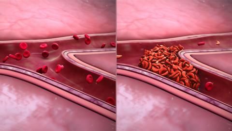
Image adpated from Scientific Animations (https://www.scientificanimations.com/wiki-images/)(, under BY-SA 4.0 License (https://creativecommons.org/licenses/by-sa/4.0/)

Redoxoma Highlights by Danilo Grünig Humberto da Silva, from Department of Biology, UNESP/IBILCE, São José do Rio Preto

Sickle cell anemia is a prevalent form of anemia caused by mutation of one aminoacid in the hemoglobin chain. This induces red blood cells (RBCs) to undergo cycles of contraction, especially under hypoxia, assuming a sickle shape, leading to impaired oxygen transport, hemolysis, vascular occlusion, profound anemia and several other clinical consequences. Redox processes have been thought to contribute to the sickling processes and their implications, but the overall pitcture remains obscure. Recent work from CEPID-Redoxoma helped unravel some of these aspects. This study investigated addressed how RBCs from patients with sickle cell anemia deal with redox metabolism disruption. It investigated the generation of specific oxidative lesions, and the levels of an unexplored antioxidant that could act as a candidate biomarker for oxidative status in sickle cell anemia. Rigorous exclusion criteria were used to obtain the studied groups, which were composed of 10 subjects without hereditary anemias and 10 sickle cell anemia patients. The results indicated that the patient´s RBCs overwhelm the antioxidant defense system, leading to an impaired antioxidant capacity that significantly contributed to the increase in lesion markers and hemolysis. Moreover, there were alterations on markers of all biochemical pathways evaluated in the patient´s RBCs. In particular, the levels of an amino acid with antioxidant properties, named ergothionein, were decreased by two-fold in patient´s RBCs. Importantly, there were strong associations of ergothioneine levels with other RBC metabolism markers, suggesting that its supplementation could be proposed as possible therapeutic alternative for sickle cell anemia treatment. Therefore, impaired antioxidant capacity results in a loss of cellular metabolism control, contributing to a decreased lifespan of the patient´s RBCs.


Article:

  • N. A. Chaves, T. G. P. Alegria, L. S. Dantas, L. E. S. Netto, S. Miyamoto, C. R. Bonini Domingos, D. G. H. da Silva. Impaired antioxidant capacity causes a disruption of metabolic homeostasis in sickle erythrocytes Free Radical Biology and Medicine, 141: 34–46, 2019. | doi: 10.1016/j.freeradbiomed.2019.05.034

Add new comment

Peroxiredoxin hyperoxidation increases in the presence of bicarbonate/carbon dioxide

Submitted by redoxoma on
peroxidase cycle, hyperoxidation pathway and interaction and oxidation of other thiol proteins, signaling

Highlights by Daniela R. Truzzi and Ohara Augusto, from Instituto de Química da USP
Corresponding author e-mail: dtruz-I-am-here-zi@hotmail.com@iq.usp.br

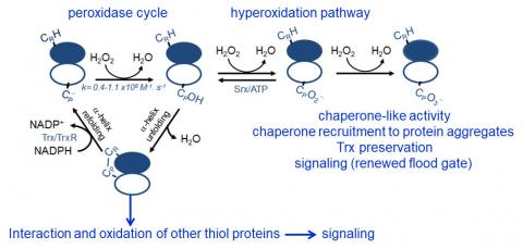
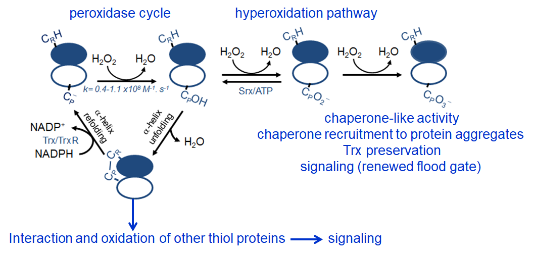
Peroxiredoxins (Prx) are abundant thiol peroxidases that react rapidly with H2O2, constituting an important antioxidant defense and acting as sensors and transmitters of H2O2 signals in cells. Eukariotic 2-Cys Prxs lose their peroxidase activity at high hydroperoxide levels in a process called hyperoxidation, which constitutes a pathway to Prx functions beyond the antioxidant activity (Fig. 1) [1]. Since the biologically ubiquitous HCO3/CO2 pair accelerates the reaction of H2O2 with several biothiols [2, 3], we examined whether physiological concentrations of the HCO3/CO2 pair (25 mM) could increase recombinant human peroxiredoxin 1 (Prx1) hyperoxidation by H2O2 [4]. Immunoblotting, kinetic and MS/MS experiments revealed that HCO3/CO2 increases Prx1 hyperoxidation and inactivation both in the presence of excess H2O2 and during enzymatic (NADPH/thioredoxin reductase/thioredoxin) and chemical (dithiothreitol) turnover. Based on previous studies, we hypothesized that the stimulating effect of HCO3/CO2 was due to HCO4 (peroxymonocarbonate), a peroxide present in equilibrated solutions of H2O2 and HCO3/CO2 [2, 3]. Indeed, additional experiments and calculations uncovered that HCO4 oxidizes CPSOH to CPSO2 with a second-order rate constant two orders of magnitude higher than H2O2 ((1.5 ± 0.1) × 10⁵ and (2.9 ± 0.2) × 10³ M⁻¹.s⁻¹, respectively) and that HCO4 is 250 times more efficient than H2O2 at inactivating 1% Prx1 per turnover [4]. The fact that the biologically ubiquitous HCO3/CO2 pair stimulates Prx1 hyperoxidation may be quite relevant to cell homeostasis because the antioxidant and redox relay functions of the enzyme decline but other actions may rise, such as the chaperone-like activity, the redox signaling pathways mediated by Cys-based proteins that are poorly reactive towards H2O2 and the maintenance of Trx-dependent activities (Fig. 1). Relevantly, parallel studies led by Christine Winterbourn and co-workers reported that the HCO3/CO2 pair also stimulates Prx2 and Prx3 hyperoxidation [5] and protein tyrosine phosphatase 1B inactivation involved in epidermal growth factormediated signaling [6]. Taking together, these recent studies confirm that HCO4 deserves further investigation as a biological oxidant [3] and point to a possible role of HCO3/CO2 levels in H2O2mediated signaling.

Peroxidase cycle, hyperoxidation pathway


Figure 1. Simplified scheme of the catalytic cycle and the hyperoxidation pathway of 2-Cys Prxs and the activities related to them.


References

  1. E. A. Veal, Z. E. Underwood, L. E. Tomalin, B. A. Morgan, C. S. Pillay. Hyperoxidation of Peroxiredoxins: Gain or Loss of Function? Antioxidants & Redox Signaling, 28(7): 574–90, 2018. | doi: 10.1089/ars.2017.7214
  2. D. F. Trindade, G. Cerchiaro, O. Augusto. A Role for Peroxymonocarbonate in the Stimulation of Biothiol Peroxidation by the Bicarbonate/Carbon Dioxide Pair Chemical Research in Toxicology, 19(11): 1475–82, 2006. | doi: 10.1021/tx060146x
  3. D. R. Truzzi, O. Augusto. Influence of CO2 on Hydroperoxide Metabolism Hydrogen Peroxide Metabolism in Health and Disease, (Vissers, M.C.M., Hampton, M., Kettle, A.J. eds) 81–99, Oxidative stress and disease, Taylor & Francis/CRC Press, Boca Raton, 2017. | doi: 10.1201/9781315154831-4
  4. D. R. Truzzi, F. R. Coelho, V. Paviani, S. V. Alves, L. E. S. Netto, O. Augusto. The bicarbonate/carbon dioxide pair increases hydrogen peroxide-mediated hyperoxidation of human peroxiredoxin 1 Journal of Biological Chemistry, 294(38): 14055–67, 2019. | doi: 10.1074/jbc.ra119.008825
  5. A. V. Peskin, P. E. Pace, C. C. Winterbourn. Enhanced hyperoxidation of peroxiredoxin 2 and peroxiredoxin 3 in the presence of bicarbonate/CO2 Free Radical Biology and Medicine, 145: 1–7, 2019. | doi: 10.1016/j.freeradbiomed.2019.09.010
  6. M. Dagnell, Q. Cheng, S. H. M. Rizvi, P. E. Pace, B. Boivin, C. C. Winterbourn, E. S. J. Arnér. Bicarbonate is essential for protein-tyrosine phosphatase 1B (PTP1B) oxidation and cellular signaling through EGF-triggered phosphorylation cascades Journal of Biological Chemistry, 294(33): 12330–8, 2019. | doi: 10.1074/jbc.ra119.009001

Add new comment

Fluorescent protein-based biosensors: potent tools to shed light on redox biochemistry and biology

Submitted by redoxoma on
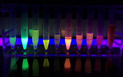
Fluorescence from fluorescent proteins excited with UV light. Picture taken by Erik A. Rodriguez. Autor: Erin Rod, licensed under CC-BY-SA 4.0
Post image by Erin Rod, via Wikimedia (licensed by CC-BY-SA 4.0)

Main Article by Marcelo A. Comini from Laboratory Redox Biology of Trypanosomes, Institut Pasteur de Montevideo, Montevideo, Uruguay.
Corresponding author e-mail: mcomin@qualqueri@pasteur.edu.uy

Thiol-redox homeostasis from a cellular viewpoint. Major cellular processes (e.g. growth, differentiation, DNA-replication, translation, metabolism, signaling and death) are redox modulated. Among the different types of redox modifications that protein residues can undergo, the reversible oxidation of the cysteine thiol group to disulfide confers this residue the potential to act as versatile molecular switch. In fact, formation or breakage of a (homo or hetero) disulfide bond may have important consequences in protein structure and conformation, and hence on its functional properties (i.e. activity, stability, interaction with partners/ligands, subcellular localization, etc.). In most organisms, thiol-disulfide homeostasis is controlled by two main redox systems: the thioredoxin system and the glutathione/glutaredoxin system; or equivalent systems in certain lineages [1, 2]. Different types of peroxidases are important components of these systems, being able to transfer their oxidation status to target proteins [3].

Location, timing and specificity are key factors for the effective maintenance of intracellular redox balance. Therefore, the redox systems are distributed in different subcellular compartments, with the oxidoreductases and peroxidases providing rapid kinetic control of the cysteine redox state of different protein targets. Worth noting, each organelle/compartment has its own redox profile [4], which implies that the intracellular redox status is not uniform but heterogeneous. This poses an important challenge for studies aimed to address from the simpler to the more complex questions in thiol-dependent redox biochemistry and biology, both at cellular and organism level.

What are the tools available for measuring thiol-disulfide redox state? A set of different chemical probes displaying specificity for thiols is available. Depending on the chemical nature of the probe, the readout of the reaction will be fluorescence, absorption or luminescence. If the protein of interest undergoes a redox-dependent conformational change that translates into a differential migration pattern under denaturing conditions, then the Western blot technique can be used to estimate the proportion of reduced and oxidized target protein. Mass spectrometry-based techniques, combined with selective thiol labeling and enrichment, offer the possibility for large scale, quantitative and high-throughput analysis of the cellular redox proteome [5]. However, taken together, these approaches present several disadvantages that can lead to erroneous conclusions. In several cases, cell integrity is destroyed because the determinations rely on extractive methods (e.g. for some chemical probes, redox Western blot or redox proteomics). This procedure demands the use of specific thiol-blocking agents to “freeze” the redox state of the proteins, in order to avoid non-specific thiol-disulfide exchange between molecules from compartments that are not in redox equilibrium. Although thiol-specific, several chemical probes are unable to distinguish protein- from low molecular weight-thiols or even be directed to specific organelles. In addition, many chemical probes react irreversibly with thiols or even have important collateral redox effects (e.g. oxidation, radical formation, cytotoxicity). In fact, without the appropriate controls (or even with them), the results, commonly referred as “overall redox status”, obtained with these methods are technically biased. Furthermore, the techniques described above provide endpoint measurements or, in other words, “a photo of the movie” that is far from representing the high dynamism that characterize cell physiology. Although multiple sampling may overcome this limitation, the experiments become laborious, prone to variability and expensive.

Can these limitations be overcome? Yes, they can and the key to unlock this door was the development of biosensor technology based on genetically-encoded fluorescent proteins (FP) [6, 7]. FP are engineered into thiol-redox biosensors by introducing a couple of vicinal cysteine residues at the protein surface and in close proximity to residues interacting with the hidden chromophore [8]. The cysteines are the biological recognition element that will “sense” and equilibrate their thiol-disulfide state with that of the surrounding environment. The modification of the redox state of these cysteines is accompanied by small and local conformational changes that modify the protonation state of the chromophore and, hence, its spectral properties [6].

A Danish group headed by Jakob R. Winther pioneered the generation and use of the first thiol-redox yellow fluorescent protein biosensor that proved useful in informing the thiol-redox status of Escherichia coli and yeast [9, 10]. Since then, a manifold of articles reported the generation of new redox sensitive FP variants that not only cover almost the full visible spectra (400-700 nm) but also were fine-tuned to operate in organelles with different redox potentials [5, 6, 7].

Depending on their photophysical properties, the FP-redox biosensors can be classified into intensiometric (single wavelength indicators that exhibit changes in their fluorescence intensity) or ratiometric (dual wavelength indicators that exhibit a shift in either their optimum absorption or emission wavelength intensities). The first are suitable for qualitative measurements because the fluorescence signal will depend not only on the concentration of the target molecule but on the expression level of the biosensor and other factors such as cell thickness and pH. The ratiometric biosensors are preferred for quantitative measurements because the interferences pointed above are cancelled out.

A major achievement in this area have been the development of biosensors coupled to different thiol-disulfide oxidoreductases (glutaredoxin, thioredoxin, mycoredoxin and bacilliredoxin) and peroxidases. Fusion to these redox active enzymes expanded the specificity for different redox species (glutathione, mycothiol, bacillithiol, proteins, and peroxides) and, due to kinetic acceleration, lowered the time scale at which redox events can be measured [e.g. from minutes to (nano)seconds].

Because they are genetically encoded, the biosensors can be expressed transiently (as episome) or stably (inducible or constitutively), the last upon integration of the reporter gene into the genome of the target cell/organism. The generation of stable cell lines allows selecting cell populations with a known and homogeneous expression level of the biosensor. At first glance, the establishment of stable redox-reporter cell lines may appear time consuming. However, it constitutes an unlimited source of material that guarantee more consistent results by bypassing the cytotoxic and epigenetic effects, and the cell-to-cell variability associated to uncontrolled transient expression-based assays.

Fluorimeter (plate reader), fluorescent microscope and flow cytometer are suited instruments to detect signals from FP biosensors. The first may be less sensitive and, hence, require a stronger expression of the reporter gene. Confocal microscopy allows for high spatiotemporal resolution at (sub)cellular level, whereas flow cytometry offers statistical robustness and high-throughput power.

With the exception of Archaea, the redox biosensors were expressed in a wide range of organisms from different Kingdoms and, so far, proved valuable to investigate fundamental questions of redox biology [6].

A bright future. The FP-based redox biosensors feature several advantages summarized in: they are produced by the cell and become functional without further intervention by the researcher and can be targeted to almost any subcellular domain/compartment wherefrom they will inform the thiol-redox state in a non-invasive, real-time and dynamic fashion. As long as there is not spectral overlap, the redox biosensors are compatible with multiparametric analysis relying on the use of complementary fluorescent probes or methods. The possibility of tagging the biosensor to different proteins opens the opportunity to “spy” redox signaling processes that control distinct cellular functions with high molecular and temporal precision. The FP biosensors can be rated as excellent biotools to address major questions in the field of redox biochemistry and biology for many pathophysiological phenomena of interest in transmissible (e.g. infectious) and non-transmissible disease’s models. Last but not least, they also show great potential in the drug discovery area, with applications that encompass the screening of drug libraries (e.g. for the selection or discard of molecules causing redox unbalance) and the study of drug toxicity [11] and mode of action [12].


References

  1. G. Salinas, M. A. Comini. Alternative Thiol-Based Redox Systems Antioxidants & Redox Signaling, 28(6): 407–9, 2018. | doi: 10.1089/ars.2017.7464
  2. C. Klomsiri, P. A. Karplus, L. B. Poole. Cysteine-Based Redox Switches in Enzymes Antioxidants & Redox Signaling, 14(6): 1065–77, 2011. | doi: 10.1089/ars.2010.3376
  3. Y. Go, D. P. Jones. Redox compartmentalization in eukaryotic cells Biochimica et Biophysica Acta (BBA) - General Subjects, 1780(11): 1273–90, 2008. | doi: 10.1016/j.bbagen.2008.01.011
  4. M. A. Comini. Measurement and meaning of cellular thiol:disufhide redox status Free Radical Research, 50(2): 246–71, 2016. | doi: 10.3109/10715762.2015.1110241
  5. A. J. Meyer, T. P. Dick. Fluorescent Protein-Based Redox Probes Antioxidants & Redox Signaling, 13(5): 621–50, 2010. | doi: 10.1089/ars.2009.2948
  6. M. Schwarzländer, T. P. Dick, A. J. Meyer, B. Morgan. Dissecting Redox Biology Using Fluorescent Protein Sensors Antioxidants & Redox Signaling, 24(13): 680–712, 2016. | doi: 10.1089/ars.2015.6266
  7. C. V. Piattoni, F. Sardi, F. Klein, S. Pantano, M. Bollati-Fogolin, M. Comini. New red-shifted fluorescent biosensor for monitoring intracellular redox changes Free Radical Biology and Medicine, 134: 545–54, 2019. | doi: 10.1016/j.freeradbiomed.2019.01.035
  8. H. Ostergaard. Shedding light on disulfide bond formation: engineering a redox switch in green fluorescent protein The EMBO Journal, 20(21): 5853–62, 2001. | doi: 10.1093/emboj/20.21.5853
  9. H. Østergaard, C. Tachibana, J. R. Winther. Monitoring disulfide bond formation in the eukaryotic cytosol The Journal of Cell Biology, 166(3): 337–45, 2004. | doi: 10.1083/jcb.200402120
  10. R. Wittig, V. Richter, S. Wittig-Blaich, P. Weber, W. S. L. Strauss, T. Bruns, T. P. Dick, H. Schneckenburger. Biosensor-Expressing Spheroid Cultures for Imaging of Drug-Induced Effects in Three Dimensions Journal of Biomolecular Screening, 18(6): 736–43, 2013. | doi: 10.1177/1087057113480525
  11. J. Franco, F. Sardi, L. Szilágyi, K. E. Kövér, K. Fehér, M. A. Comini. Diglycosyl diselenides alter redox homeostasis and glucose consumption of infective African trypanosomes International Journal for Parasitology: Drugs and Drug Resistance, 7(3): 303–13, 2017. | doi: 10.1016/j.ijpddr.2017.08.001

Marcelo Comini (mcomini@pasteur.edu.uy) holds a degree in Biochemistry (1999) from the Universidad Nacional del Litoral (Santa Fe, Argentina) and a Dr. rerum naturarum (2004) from the Technical University of Braunschweig (Braunschweig, Germany), with a postdoctoral period (2004-2007) at the Biochemistry Centre from the Heidelberg University (Heidelberg, Germany). In 2008, he joined the Institut Pasteur de Montevideo as “Young group leader” of the Laboratory Redox Biology of Trypanosomes and five years later became Principal Investigator. He is a foundational member of the Molecular, Cellular and Animal Technology Program (ProTeMCA) from IP-Montevideo. He was invited to join several European and International consortia devoted to drug discovery research against neglected diseases. He is member of the Sistema Nacional de Investigadores (level 2) and Programa de Desarrollo de las Ciencias Básicas (level 4) from Uruguay. His scientific contributions (59 research articles and 9 book chapters) were published in top peer-reviewed journals and books. The research interest of his team embraces: understanding fundamental aspects of the redox biology of trypanosomatids, the early phase of drug discovery against these pathogens and the development of fluorescent protein-based biosensors.

Add new comment

The "D" of CEPID

Submitted by redoxoma on
"D"

Education Page by Carmen Fernandez
Corresponding author e-mail: carmecarmeqn@hotmail.com@iq.usp.br

The most important attribute of the special program CEPID [1] (Centros de Pesquisa, Inovação e Difusão, or RIDC, Research, Innovation, and Dissemination Centers) is the multiplicity of its missions. Thus, in addition to the primary mission of developing research on specific themes, CEPID´s are characterized by the development of effective mechanisms of Technology Transfer and Education and Knowledge Diffusion. In this article, we focus on the mission of Education and Diffusion of Knowledge of Redoxoma [2], translated by D from CEPID. A careful reading of Fapesp's 2011 initial public announcement [3] reveals that what is expected in D encompasses quite diverse actions such as:

[...] centers must maintain diffusion activities and contribute to the improvement of science education; [...] they should contribute to the scientific education of children and young people, in the activity of scientific diffusion, be it interacting with schools or interacting directly with the general public; [...] in addition to performing extension activities in the area of basic education, such as scientific initiation for students and teachers of high school, teacher training and courses of scientific diffusion.

Difficult to translate all this into a simple D. In fact, what is intended are educational actions, both in the contact with students of basic education and with the general public, promotion of professional development of teachers in addition to actions to raise public understanding of science. Apart from the question of covering several actions in D, it is worth taking advantage of this space to address the issues of ambiguities that usually appear with the terms Diffusion / Dissemination. And in the sequence we will deal with the D actions of Redoxoma. Difusão incorporates Divulgação científica, Disseminação científica and Jornalismo científico itself

The D in the English version of the Fapesp website [1] is treated as Dissemination and in the Portuguese version it is translated as Difusão (Diffusion). Beyond the different words for the English terms - diffusion, dissemination, public awareness of Science (PAwS), public understanding of science (PUS), or more recently, Public Engagement with Science and Technology (PEST), among others, there are also ambiguities of the terms in the Portuguese language - the terms Difusão, Disseminação and Divulgação are often used as synonyms. So it is worth treating these differences of the terms in Portuguese, although this text is written in English. In an attempt to make it clearer we will use the Portuguese terms in italics to avoid generating more misunderstandings.

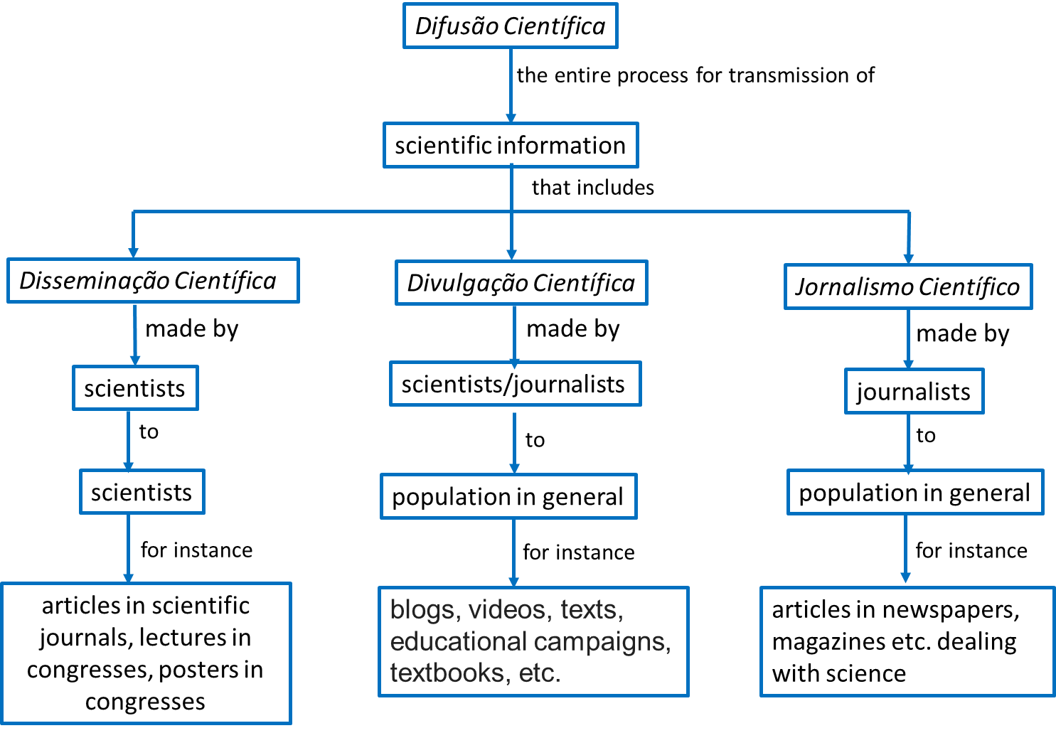
According to Bueno [4] Difusão is the broader term and refers to any and all processes or resources used for communicating scientific information. Thus, Difusão incorporates Divulgação científica, Disseminação científica and Jornalismo científico itself (Figure 1). What changes is the target audience and therefore the level of language used. If the Difusão is made for experts, it is called Disseminação científica and occurs with specific technical terms of a given area (for example it is what occurs in conferences and scientific articles), if it is made for the general public it is called Divulgação científica (communicating science to public understanding). The latter presupposes a recoding of the specialized language of the experts into a language comprehensible to a non-specialist audience [5], for instance, videos, texts, explanatory health campaigns, textbooks, documentaries, etc. Also, there is the term Jornalismo científico (Science journalism) that is used when the article dealing with science is written by a journalist.

Difusão Científica

Figure 1. Differences between the terms Difusão, Divulgação, Disseminação Científica and Jornalismo Científico.

Thus, in D of CEPID several activities are being included as the Disseminação científica (represented by the articles and lectures of scientists for public experts and also, this newsletter [6] that is another channel for the dissemination of Redoxoma researches), Divulgação científica (to students and non-specialized audiences), and Jornalismo científico (from articles produced for the Redoxoma website) as well as several other actions related to basic education and teacher training activities. And in this range of activities under the letter D from our CEPID Redoxoma, are also included activities of educational research that feedback our Divulgação actions.

Now we will focus specifically on the D actions of Redoxoma. It is noteworthy that the research of CEPID Redoxoma focuses on the redox mechanisms involved in cellular homeostasis and pathologies, with emphasis on chronic-degenerative diseases, such as cardiovascular, metabolic and inflammatory diseases. Thinking about the Difusão actions students have problems in associating the concept of oxireduction reactions with the context of the day to day and the contact with the basic education and the training of teachers, the chemical content that connects all of this is oxidation reactions or more generally speaking, redox processes.

In this sense, the approach of our group is to connect all these activities - educational research, teacher training, and Difusão Científica. It starts from educational research to investigate the difficulties with understanding the redox subject at all levels in order to adopt the best strategies, resources, and language, and we arrive at the development of proposals that take into account the difficulties with this subject. The activities developed are a backdrop for further educational research to be carried out.

Speaking briefly of the difficulties with this subject, students have problems in associating the concept of oxireduction reactions with the context of the day to day, in spite of the importance of the redox processes and their daily applications. On the other hand, some teachers find the subject difficult to teach, and because of this, they feel that lesson plans are difficult to prepare. In chemistry teaching in general and in teaching redox processes in particular, students have many difficulties in traffic that scientists easily make between the macroscopic (the phenomenon), submicroscopic (particles and their arrangement) and symbolic levels (formulas, chemical equations, signal charges, etc.), see figure 2. In addition, there are problems with the coexistence of several explanatory models, with the understanding of the simultaneity of oxidation and reduction reactions, with a confusion between the language used in physics for electrical circuits and that of chemistry when dealing with cells and batteries, among many others.

Difusão Científica

Figure 2. Johnstone's model of the three levels of Chemical Knowledge [7].

Our group has developed several analyzes with the teaching of redox processes and brought this knowledge to Divulgação actions, production of didactic resources, courses for teachers and content for the media. Some of the group actions will be mentioned here. We analyzed the difficulties with the school redox content in the Brazilian context and analyzed textbooks approved by the federal government and used in all basic education (from the first to the ninth year of elementary school and in the three years of high school). Subjects such as cellular respiration, photosynthesis, cells and batteries, rust and combustion already appear in the first years of the elementary school in a more contextualized and phenomenological way and are increasingly being formalized and translated into a submicroscopic and symbolic level in High School. The visual representations in the books were a specific theme of analysis and what is observed is an excessive valuation in the books of representations at the macro level (mainly pictures) and in much smaller quantity appear the representations at the submicroscopic and symbolic level (Figure 3). And still in smaller numbers appear representations in which the three different levels of the chemistry are contemplated. There are no explanations on how to move between these different levels and this is a task that is left to the teacher. It turns out that we have also analyzed teacher's classes in this aspect and what we observe is that the teacher transits between these levels as if it were obvious to his students. In the textbooks are perceived representations and texts that end up emphasizing still more the difficulties of the students. The group has also produced didactic resources that take into account the difficulties pointed out. Thus, simulators, videos, and games were produced and made available on the Labiq [8] website. The group also developed and offered several courses for teachers that showed this repertoire of difficulties, textbooks, and student resources.

(A) Macroscopic level, (B) Submicroscopic level, (C) Macroscopic and Symbolic level

Figure 3. Examples of textbook representations at (A) Macroscopic level, (B) Submicroscopic level, (C) Macroscopic and Symbolic level.
Santos, W. L. and Mól, G. S. (2013). Química Cidadã (2ed. Vol.3). São Paulo: AJS Ltda. Reproduced by permission of AJS Ltda, São Paulo, São Paulo.

In terms of Divulgação, Redoxoma has regularly participated in lecture programs in public places intended for general audiences. Examples are: Pint of Science [9] (presentations of scientists in bars), Chemistry is life (lectures by scientists at the Mário de Andrade Library [10]), Chemistry Week [11] at IQ-USP, 24 hours of Science [12], USP Science and Technology Week [13], National Science and Technology Week [14], among others. An exhibition was produced for the São Paulo subway on Aging and was developed a site named Free and Radicals [2,15] with explanatory material. Content was also produced for the media (Jornalismo científico) where research activities related to technology transfer and education and Divulgação científica are made available to the public through articles published on the Redoxoma´s website [2], shared on the Facebook page [16] and distributed as a press release for Fapesp Agency [17] and the Journal of USP [18]. This material generated new reports and videos, expanding the Divulgação of Redoxoma.

In terms of training of people for the Chemical Education area, six masters dissertations and nine doctoral theses were defended in the group since the project start (2012) and three master's degrees and nine doctorates are in progress. Lists of these surveys and publications are presented on the Redoxoma website in the annual report tab [19], Labiq [8] and PEQuim (Pesquisa em Ensino de Química, Research in Chemistry Teaching) group’s page [20].


References

  1. FAPESP. Centros de Pesquisa, Inovação e Difusão (Homepage)url: http://cepid.fapesp.br/home/
  2. CEPID Redoxoma. Centro de Pesquisa em Processos Redox em Biomedicina (Homepage)url: http://redoxoma.iq.usp.br/
  3. FAPESP. Edital CEPID 2011url: http://www.fapesp.br/6335
  4. W. da C. Bueno. Jornalismos científico: coneceitos e funções Ciência e Cultura, 9(37): 1420-1428, 1985.url: http://www.scielo.br/scielo.php?script
  5. J. M. M. Loureiro. Museu de ciência, divulgação científica e hegemonia Ciência da Informação, 32(1): 88–95, 2003. | doi: 10.1590/s0100-19652003000100009
  6. CEPID Redoxoma. Redoxoma Newsletter (Homepage)url: http://redoxomanewsletter.iq.usp.br/
  7. A. H. Johnstone. Why is science difficult to learn? Things are seldom what they seem Journal of Computer Assisted Learning, 7(2): 75–83, 1991. | doi: 10.1111/j.1365-2729.1991.tb00230.x
  8. LABIQ. Laboratório Integrado de Química e Bioquímica (Homepage)url: http://labiq.iq.usp.br/
  9. Pint of Science. Pint of Science Brasil (Homepage)url: https://pintofscience.com.br/
  10. Prefeitura de SP. Biblioteca Mário de Andrade (Homepage)url: https://www.prefeitura.sp.gov.br/cidade/secretarias/cultura/bma/
  11. Instituto de Química - USP. Semana da Química (Homepage)url: http://www.iq.usp.br/semanadaquimica/
  12. USP. Virada Científica (Homepage)url: https://www5.usp.br/tag/virada-cientifica/
  13. PRCEU-USP. Semana USP de Ciência e Tecnologia 2018 (Homepage)url: http://prceu.usp.br/noticia/ciencia-e-tecnologia-na-usp-2018/
  14. MCTIC. Semana Nacional de Ciência e Tecnologia 2019 (Homepage)url: http://snct.mctic.gov.br/portal
  15. CEPID Redoxoma. Livres e Radicais: Química, vida, saúde e radicais livres (Homepage)url: http://livresradicais.iq.usp.br/
  16. CEPID Redoxoma. Centro de Pesquisa em Processos Redox em Biomedicina (Canal do Facebook)url: https://www.facebook.com/redoxoma/
  17. FAPESP. Agência Fapesp (Homepage)url: http://agencia.fapesp.br/inicial/
  18. USP. Jornal da USP (Homepage)url: https://jornal.usp.br/
  19. CEPID Redoxoma. Relatório Anual de Atividades (CEPID Redoxoma),url: http://redoxoma.iq.usp.br/paginas_view.php?idPagina
  20. C. Fernandez. PEQuim - Grupo de Pesquisa em Ensino de Química - USP (Homepage)url: https://sites.usp.br/pequim/

Carmen Fernandez, Ph.D. Associate Professor at Department of Fundamental Chemistry,
Institute of Chemistry, University of São Paulo, Brazil


Add new comment

A Rank of the Most Popular Redox Genes

Submitted by redoxoma on
A Rank of the Most Popular Redox Genes

Redoxoma Highlights by Isaias Glezer

The obsession for ranks of top something or someone is not simple to explain, but one must admit their influence. This has been highlighted in an editorial commenta focusing on a list released by Natureb of the 100 most-cited scientific papers. Curiously or not, the top most-cited paper is a protein quantification method developed by Lowry [1]. Rankings can be indeed useful to assess and compare achievements. One year ago, Nature published a News feature article entitled ‘The most popular genes in the human genome’ [2]⁠, which revisited a similar 2014 ranking. The top-10 list is significantly dominated by cancer-related genes, leaded by the tumor repressor TP53 with 8,954 citations.c I was eager to know what would be the rank of redox-associated genes and whether this list would bring interesting insights and surprises. I decided to rank redox genes according to four different organisms, namely human, mouse, rat and yeast, as explained below.

Notes
  1. www.sciencemag.org
  2. www.nature.com
  3. from 1990 to 2017/12/31 (slightly updated, and for this reason, higher than reported in Nature). Scripts developed by Peter Kerpedjiev and available at github.com.
  4. GO:0016491 and GO:0016209, evidences: experimental and manual sequence orthology.
  5. enrichR package for R environment, online available

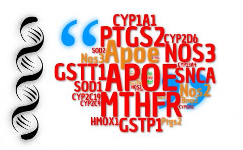
The rationale of this ranking was to identify articles (citations) that describe some information on what a specific gene does using US National Library of Medicine (NLM) data. For each gene, publication counts were aggregated over the years, according to previously reported strategies.c Organisms were evaluated individually, and each list was filtered and re-ranked exclusively for redox genes. Here, a redox gene list was generated by combining items classified by molecular function gene ontology (GO) as oxidoreductase or antioxidant activity.d In humans, APOE gene tops the redox list with 4,116 citations (Fig. 1A). Apolipoprotein E is mostly studied for its role on lipid metabolism and as one of the mostly established risk factors for Alzheimer Disease along with age (allele e4). Why APOE was included in a redox gene list? The answer comes from its classification as antioxidant molecular function. In fact, it has been shown that APOE protects cells against H2O2-induced oxidative damage, possibly by a metal binding mechanism [3]⁠. Methylenetetrahydrofolate reductase (MTHFR) is in second place. Mutations in this gene have been associated with vascular disease due to hyperhomocysteinaemia [4]⁠, and deficiency of this enzyme affects other conditions relevant to human health. SNCA (ranked 6th), which encode α-synuclein, is implicated in Parkinson Disease as the major component of intracellular Lewy body inclusions in affected dopaminergic neurons. A redox activity of α-synuclein-Cu(II) has been reported [5]⁠. Maybe, none of these three genes would be mentioned as top representative examples of redox biology molecules by an expert, although the mentioned diseases have appeal for this field of research and MTHFR is implicated in methionine recicling and transsulfuration pathway . Apart from fashionable biases, should genes like these be more explored in redox chemistry and biology? Or is it timely that a more ellaborated list of meaningful redox genes is performed? These questions become more important with the exponential growth of functional genomics studies during the last decade (see Figure 2 for a timeline).

Figure 1

Figure 1 – Top 20 genes according to citations (articles describing gene functions and features) for a complete ranking (left) and redox genes ranking (right; hashtag indicates original position in the complete ranking). The panel depicts human (A), murine (B), rat (D) and yeast (D) gene lists.


Figure 2 – Citations of the top 10 redox genes over de last 20 years (1997 – 2017).

Research on inflammation has also driven the investigation of genes encoding enzymes involved in the generation of signaling molecules from electron transfer reactions that produce endogenous free radicals. As examples, the synthesis of prostanoids and superoxide radical by cyclooxygenase II (PTGS2; 3rd in human - 4th in mouse and 2nd in rat), and nitric oxide by NOS2 (16th in human, 2nd in mouse and 1st in rat). In vascular research, endothelial nitric oxide synthase (NOS3; 4th in human, 3rd in mouse and rat) highlights the historical importance of this free radical in human (patho)physiology. It is also interesting that mouse and rat gene lists depict a more clear scenario vs. human of the redox context in biomedical research, attesting the importance of genes that supported landmark discoveries in the field (Sod1/2, Homx1, Cat, Cybb, Nqo1, Nox4, Gpx1, etc.) [6]⁠. SOD1 also tops the list for yeast redox genes (2nd), only surpassed by a less typical redox gene, URE2 (1st), which possesses glutathione peroxidase activity [7]⁠. However, URE2 protein product is mostly characterized for nitrogen metabolism and a prion-like behavior. Also, in terms of marginal redox appeal, the top list for human presents several genes encoding components of cytochrome P450 involved in xenobiotic, prostanoid and steroid metabolization (CYP1.../2.../3…). Although the chemical reactions involved are clearly redox [8]⁠, the focus of gene function investigation relate to other research fields (pharmacokinetics, endocrinology, etc.). Glutathione S-Transferase Theta 1 (GSTT1, 5th in human list) is involved in drug metabolism, and is also a candidate gene for cancer risk along with GSTP1 (7th). GSTs are well known detoxifying enzymes induced by oxidative stress [9]⁠. Submitting the putative top100 redox genes to enrichmente analysis revealed predominance of hydrogen peroxide metabolic processes and compartmentalization to the mitochondria (Fig. 3) Checking the ranking for human redox genes, not surprisingly arachidonic acid and steroid metabolism showed predominant enrichment, while interestingly cellular response to oxidative stress ranked slightly below those processes.


Figure 3 – The list of top 100 human redox genes according to GOs Biological Process and Cellular Component (2018 classification).

Overall, the top lists of redox genes can partially fulfill our expectations, or raise questions whether some popular genes from early 90’s to 2017 should be considered representative redox components or not. A complete ranking can also reveal which genes have been neglected or not yet explored. This brief discussion attempts to call attention to our current classification and investigation focus on redox genes, and if we could call them as such. Perhaps a tentative conclusion is that a better discrimination and classification of gene ontologies for redox genes is needed and timely.


References

  1. O. H. Lowry, N. J. Rosebrough, R. J. Farr. Protein measurement with the Folin phenol reagent Journal of Biological Chemistry, 193(1): 265–75, 1951 | view paper
  2. E. Dolgin. The most popular genes in the human genome Nature, 551(7681): 427–31, 2017 | doi: 10.1038/d41586-017-07291-9
  3. M. Miyata, J. D. Smith. Apolipoprotein E allele–specific antioxidant activity and effects on cytotoxicity by oxidative insults and β–amyloid peptides Nature Genetics, 14(1): 55–61, 1996 | doi: 10.1038/ng0996-55
  4. P. Frosst, H. Blom, R. Milos, P. Goyette, C. Sheppard, R. Matthews, G. Boers, M. den Heijer, L. Kluijtmans, L. van den Heuve, e. al. et al.. A candidate genetic risk factor for vascular disease: a common mutation in methylenetetrahydrofolate reductase Nature Genetics, 10(1): 111–3, 1995 | doi: 10.1038/ng0595-111
  5. G. Meloni, M. Vašák. Redox activity of α-synuclein–Cu is silenced by Zn7-metallothionein-3 Free Radical Biology and Medicine, 50(11): 1471–9, 2011 | doi: 10.1016/j.freeradbiomed.2011.02.003
  6. W. Dröge. Free Radicals in the Physiological Control of Cell Function Physiological Reviews, 82(1): 47–95, 2002 | doi: 10.1152/physrev.00018.2001
  7. M. Bai, J. Zhou, S. Perrett. The Yeast Prion Protein Ure2 Shows Glutathione Peroxidase Activity in Both Native and Fibrillar Forms Journal of Biological Chemistry, 279(48): 50025–30, 2004 | doi: 10.1074/jbc.m406612200
  8. O. Augusto, H. S. Beilan, P. R. Ortiz de Montellano. The Catalytic Mechanism of Cytochrome P-450 Journal of Biological Chemistry, 257(19): 11288–95, 1982 | view paper
  9. J. D. Hayes, D. J. Pulford. The Glut athione S-Transferase Supergene Family: Regulation of GST and the Contribution of the lsoenzymes to Cancer Chemoprotection and Drug Resistance Part II Critical Reviews in Biochemistry and Molecular Biology, 30(6): 521–600, 1995 | doi: 10.3109/10409239509083492

Isaias Glezer, Ph.D. Assistant Professor at Department of Biochemistry,
Universidade Federal de São Paulo/Escola Paulista de Medicina


Add new comment