A Rank of the Most Popular Redox Genes

Redoxoma Highlights by Isaias Glezer
The obsession for ranks of top something or someone is not simple to explain, but one must admit their influence. This has been highlighted in an editorial commenta focusing on a list released by Natureb of the 100 most-cited scientific papers. Curiously or not, the top most-cited paper is a protein quantification method developed by Lowry [1]. Rankings can be indeed useful to assess and compare achievements. One year ago, Nature published a News feature article entitled ‘The most popular genes in the human genome’ [2], which revisited a similar 2014 ranking. The top-10 list is significantly dominated by cancer-related genes, leaded by the tumor repressor TP53 with 8,954 citations.c I was eager to know what would be the rank of redox-associated genes and whether this list would bring interesting insights and surprises. I decided to rank redox genes according to four different organisms, namely human, mouse, rat and yeast, as explained below.
- www.sciencemag.org
- www.nature.com
- from 1990 to 2017/12/31 (slightly updated, and for this reason, higher than reported in Nature). Scripts developed by Peter Kerpedjiev and available at github.com.
- GO:0016491 and GO:0016209, evidences: experimental and manual sequence orthology.
- enrichR package for R environment, online available
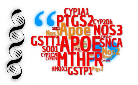
The rationale of this ranking was to identify articles (citations) that describe some information on what a specific gene does using US National Library of Medicine (NLM) data. For each gene, publication counts were aggregated over the years, according to previously reported strategies.c Organisms were evaluated individually, and each list was filtered and re-ranked exclusively for redox genes. Here, a redox gene list was generated by combining items classified by molecular function gene ontology (GO) as oxidoreductase or antioxidant activity.d In humans, APOE gene tops the redox list with 4,116 citations (Fig. 1A). Apolipoprotein E is mostly studied for its role on lipid metabolism and as one of the mostly established risk factors for Alzheimer Disease along with age (allele e4). Why APOE was included in a redox gene list? The answer comes from its classification as antioxidant molecular function. In fact, it has been shown that APOE protects cells against H2O2-induced oxidative damage, possibly by a metal binding mechanism [3]. Methylenetetrahydrofolate reductase (MTHFR) is in second place. Mutations in this gene have been associated with vascular disease due to hyperhomocysteinaemia [4], and deficiency of this enzyme affects other conditions relevant to human health. SNCA (ranked 6th), which encode α-synuclein, is implicated in Parkinson Disease as the major component of intracellular Lewy body inclusions in affected dopaminergic neurons. A redox activity of α-synuclein-Cu(II) has been reported [5]. Maybe, none of these three genes would be mentioned as top representative examples of redox biology molecules by an expert, although the mentioned diseases have appeal for this field of research and MTHFR is implicated in methionine recicling and transsulfuration pathway . Apart from fashionable biases, should genes like these be more explored in redox chemistry and biology? Or is it timely that a more ellaborated list of meaningful redox genes is performed? These questions become more important with the exponential growth of functional genomics studies during the last decade (see Figure 2 for a timeline).
Figure 1 – Top 20 genes according to citations (articles describing gene functions and features) for a complete ranking (left) and redox genes ranking (right; hashtag indicates original position in the complete ranking). The panel depicts human (A), murine (B), rat (D) and yeast (D) gene lists.
Research on inflammation has also driven the investigation of genes encoding enzymes involved in the generation of signaling molecules from electron transfer reactions that produce endogenous free radicals. As examples, the synthesis of prostanoids and superoxide radical by cyclooxygenase II (PTGS2; 3rd in human - 4th in mouse and 2nd in rat), and nitric oxide by NOS2 (16th in human, 2nd in mouse and 1st in rat). In vascular research, endothelial nitric oxide synthase (NOS3; 4th in human, 3rd in mouse and rat) highlights the historical importance of this free radical in human (patho)physiology. It is also interesting that mouse and rat gene lists depict a more clear scenario vs. human of the redox context in biomedical research, attesting the importance of genes that supported landmark discoveries in the field (Sod1/2, Homx1, Cat, Cybb, Nqo1, Nox4, Gpx1, etc.) [6]. SOD1 also tops the list for yeast redox genes (2nd), only surpassed by a less typical redox gene, URE2 (1st), which possesses glutathione peroxidase activity [7]. However, URE2 protein product is mostly characterized for nitrogen metabolism and a prion-like behavior. Also, in terms of marginal redox appeal, the top list for human presents several genes encoding components of cytochrome P450 involved in xenobiotic, prostanoid and steroid metabolization (CYP1.../2.../3…). Although the chemical reactions involved are clearly redox [8], the focus of gene function investigation relate to other research fields (pharmacokinetics, endocrinology, etc.). Glutathione S-Transferase Theta 1 (GSTT1, 5th in human list) is involved in drug metabolism, and is also a candidate gene for cancer risk along with GSTP1 (7th). GSTs are well known detoxifying enzymes induced by oxidative stress [9]. Submitting the putative top100 redox genes to enrichmente analysis revealed predominance of hydrogen peroxide metabolic processes and compartmentalization to the mitochondria (Fig. 3) Checking the ranking for human redox genes, not surprisingly arachidonic acid and steroid metabolism showed predominant enrichment, while interestingly cellular response to oxidative stress ranked slightly below those processes.
Figure 3 – The list of top 100 human redox genes according to GOs Biological Process and Cellular Component (2018 classification).
Overall, the top lists of redox genes can partially fulfill our expectations, or raise questions whether some popular genes from early 90’s to 2017 should be considered representative redox components or not. A complete ranking can also reveal which genes have been neglected or not yet explored. This brief discussion attempts to call attention to our current classification and investigation focus on redox genes, and if we could call them as such. Perhaps a tentative conclusion is that a better discrimination and classification of gene ontologies for redox genes is needed and timely.
References
- O. H. Lowry, N. J. Rosebrough, R. J. Farr. Protein measurement with the Folin phenol reagent Journal of Biological Chemistry, 193(1): 265–75, 1951 | view paper
- E. Dolgin. The most popular genes in the human genome Nature, 551(7681): 427–31, 2017 | doi: 10.1038/d41586-017-07291-9
- M. Miyata, J. D. Smith. Apolipoprotein E allele–specific antioxidant activity and effects on cytotoxicity by oxidative insults and β–amyloid peptides Nature Genetics, 14(1): 55–61, 1996 | doi: 10.1038/ng0996-55
- P. Frosst, H. Blom, R. Milos, P. Goyette, C. Sheppard, R. Matthews, G. Boers, M. den Heijer, L. Kluijtmans, L. van den Heuve, e. al. et al.. A candidate genetic risk factor for vascular disease: a common mutation in methylenetetrahydrofolate reductase Nature Genetics, 10(1): 111–3, 1995 | doi: 10.1038/ng0595-111
- G. Meloni, M. Vašák. Redox activity of α-synuclein–Cu is silenced by Zn7-metallothionein-3 Free Radical Biology and Medicine, 50(11): 1471–9, 2011 | doi: 10.1016/j.freeradbiomed.2011.02.003
- W. Dröge. Free Radicals in the Physiological Control of Cell Function Physiological Reviews, 82(1): 47–95, 2002 | doi: 10.1152/physrev.00018.2001
- M. Bai, J. Zhou, S. Perrett. The Yeast Prion Protein Ure2 Shows Glutathione Peroxidase Activity in Both Native and Fibrillar Forms Journal of Biological Chemistry, 279(48): 50025–30, 2004 | doi: 10.1074/jbc.m406612200
- O. Augusto, H. S. Beilan, P. R. Ortiz de Montellano. The Catalytic Mechanism of Cytochrome P-450 Journal of Biological Chemistry, 257(19): 11288–95, 1982 | view paper
- J. D. Hayes, D. J. Pulford. The Glut athione S-Transferase Supergene Family: Regulation of GST and the Contribution of the lsoenzymes to Cancer Chemoprotection and Drug Resistance Part II Critical Reviews in Biochemistry and Molecular Biology, 30(6): 521–600, 1995 | doi: 10.3109/10409239509083492
Isaias Glezer, Ph.D. Assistant Professor at Department of Biochemistry,
Universidade Federal de São Paulo/Escola Paulista de Medicina

Add new comment