
cells

Neutrophils add another layer of complexity to redox signaling

Submitted by redoxoma on Wed, 05/15/2019 - 18:52
neutrophil

Redoxoma Highlights by Luiz Felipe de Souza

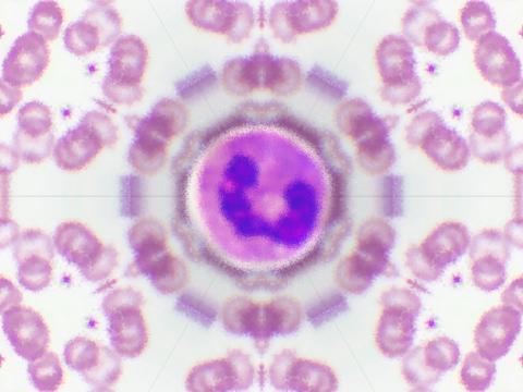
Neutrophils kill invading microbes with a myriad of antimicrobial agents and powerful oxidants. Phagocytosis and other stimuli activate the NADPH oxidase complex that generates high amounts of oxidants, which are crucial to host defense but can also promote damage to host tissue and jeopardize neutrophil function. neutrophils were traditionally considered to be a “kamikaze” cell Thus, neutrophils were traditionally considered to be a “kamikaze” cell, dying quickly after activation due to self-inflicted oxidative damage. However, it is becoming clear that neutrophils can survive for several hours after activation and are important both for regulation and termination of inflammation by releasing cytokines and other inflammatory mediators. This suggests that neutrophils must have highly efficient antioxidant systems to protect ongoing functions. We examined this hypothesis by studying peroxiredoxin in these cells [1].

Peroxiredoxins (Prxs) are thiol peroxidases that are particularly sensitive to oxidation. Prxs rely on highly reactive cysteine thiols for their antioxidant functions. Because of their high abundance and fast reactivity, Prxs are thought to act as peroxide sensors, normally being in their reduced form, and becoming oxidized as the environment shifts towards a more oxidizing state [2]. This holds true for most mammalian cells, but this system seems to be more complicated in neutrophils. By analyzing the redox state of the cytoplasmic Prxs (Prx1 and 2) in non-stimulated neutrophils, we detected that almost all the Prx pool was already oxidized in these conditions [1]. Prxs of other lymphocytes taken from the same blood showed only about 15-20% of Prx oxidation, indicating that this phenomenon might be unique to neutrophils. Inhibition of the NAPDH oxidase complex or stimulation of oxidative burst did not change the oxidative state of Prx, suggesting that the recycling system may be deficient in neutrophils. Most puzzling, thioredoxin and thioredoxin reductase, the two enzymes directly responsible for Prx recycling, were expressed at reasonable levels in neutrophils, and in contrast to Prx, thioredoxin was mainly reduced in these cells. In contrast, Prxs in acute promyelocytic leukemia HL-60 cells induced to differentiate towards a neutrophil-like phenotype were reduced under non-stimulated conditions and became oxidized upon phagocytosis. Intriguingly, differentiation of the HL-60 cells by both retinoic acid and dimethyl sulfoxide let to a robust down-regulation of Prx1, indicating that this protein may play a role in the differentiation process [1].

These data raise a number of questions regarding the redox metabolism of neutrophils. It is known that oxidation of Prxs can control cell signaling by a disulfide relay mechanism. For example, Prx1 oxidation can activate apoptosis by promoting the oxidation of ASK1, and Prx2 can oxidize and repress STAT3 signaling [3,4]. So, how are these systems operating inside the neutrophils? And why does Prxs appear “locked” as disulfides when other thiols such as thioredoxin and glutathione are reduced? Perhaps trying to answer these questions may bring other exciting findings in the field of redox research.


References

  1. L. F. de Souza, A. G. Pearson, P. E. Pace, A. L. Dafre, M. B. Hampton, F. C. Meotti, C. C. Winterbourn. Peroxiredoxin expression and redox status in neutrophils and HL-60 cells Free Radical Biology and Medicine, 135: 227–34, 2019. | doi: 10.1016/j.freeradbiomed.2019.03.007
  2. R. A. Poynton, M. B. Hampton. Peroxiredoxins as biomarkers of oxidative stress Biochimica et Biophysica Acta (BBA) - General Subjects, 1840(2): 906–12, 2014. | doi: 10.1016/j.bbagen.2013.08.001
  3. R. M. Jarvis, S. M. Hughes, E. C. Ledgerwood. Peroxiredoxin 1 functions as a signal peroxidase to receive, transduce, and transmit peroxide signals in mammalian cells Free Radical Biology and Medicine, 53(7): 1522–30, 2012. | doi: 10.1016/j.freeradbiomed.2012.08.001
  4. M. C. Sobotta, W. Liou, S. Stöcker, D. Talwar, M. Oehler, T. Ruppert, A. N. D. Scharf, T. P. Dick. Peroxiredoxin-2 and STAT3 form a redox relay for H2O2 signaling Nature Chemical Biology, 11(1): 64–70, 2014. | doi: 10.1038/nchembio.1695

Luiz Felipe de Souza, Pos-doc at Laboratory of Redox Processes in Inflammation at Department of Biochemistry,
Institute of Chemistry, University of São Paulo, Brazil


Add new comment

A molecular pathway for Protein disulfide isomerase effects on vascular Nox1-dependent oxidant generation

Submitted by redoxoma on Thu, 02/28/2019 - 14:43
Cardiovascular disease

Redoxoma Highlights by Lucia R. Lopes

Superoxide and hydrogen peroxide are important signaling intermediates in cell physiology and local imbalances in their production/removal rates promote oxidative stress, which is a major contributing factor for cardiovascular disease. The primary enzymatic sources of superoxide/hydrogen peroxide with signaling purposes in vascular cells are the Nox (NADPH oxidase) family complexes. Nox1 isoform constitutes a catalytic core at the cell membrane and is constitutively activated in cells (e.g. vascular cells) upon the expression of its canonical regulatory subunits NoxO1 (organizing subunit 1) and NoxA1 (Nox activating subunit 1) [1]. In contrast, p47phox and p67phox homologous to NoxO1 and NoxA1, respectively allow for inducible activation of Nox1 [2]. This is dependent on the phosphorylation of p47phox which produces its conformational change allowing its binding to p22phox and p67phox , culminating in Nox1 activation. Understanding mechanisms whereby Nox catalytic and regulatory subunits are assembled and their expression modulated in physiological or pathological scenarios is a major goal of redox biomedicine.

Protein disulfide isomerase (PDI) belongs to the thioredoxin superfamily of dithiol-disulfide oxidoreductases and its canonical function is to catalyze the correct insertion of disulfide bonds in nascent proteins at the endoplasmic reticulum. Previous work developed at our Redoxoma groups have shown that PDI exerts a critical role in Nox regulation, being essential for growth factor-dependent activation and expression of Nox1, as well as for Nox1-dependent vascular cell migration [3]. However, the mechanisms whereby PDI regulate Nox1 have remained unclear. In a collaborative study with Prof. Francis Miller from Duke University [4], we have now provided a compelling molecular mechanism for PDI-mediated Nox1 regulation. In a series of experiments, we showed that the Nox subunit p47phox is a relevant target of PDI effects. In vitro experiments with recombinant enzymes and mass spectrometry techniques showed that PDI physically interacts with p47phox, forming cross-linked disulfides, an interaction that facilitates p47phox phosphorylation and ensuing Nox1 activation. Cysteine 196 in p47phox and cysteine 400 at the redox active site of PDI are essential for the activation of Nox1 by PDI. Transfection of vascular smooth muscle cells (VSMC) with those proteins mutated in such critical cysteine residues prevented Nox1 activation and VSMC migration in response to the growth factor/vasoconstrictor agonist angiotensin II. Such effects were similar to those described for inhibition/silencing of PDI. Further experiments indicated that mechanisms whereby PDI enhance p47phox phosphorylation involve a reduction of an intramolecular disulfide bond between cys196 and 378 in p47phox, allowing further phosphorylation of the protein and increasing the activation of Nox1. Our findings of increased PDI and Nox1 expression in atherosclerotic aortae and of PDI interaction with p47phox in injured arteries suggest that PDI interaction with p47phox is a relevant mechanism for the effects of PDI as a redox activator of Nox1 in the vasculature. Accordingly, we found a positive correlation between the expression of PDI and p47phox in human atheroma plaques. Altogether, these in vivo data indicate that PDI interaction with p47phox contributes to oxidant generation in contexts related to atherosclerosis 4. Such a novel interaction may provide potential therapeutic targets against vascular disease.

Normal -> Atherosclerosis

References

  1. B. Bánfi, R. A. Clark, K. Steger, K. Krause. Two Novel Proteins Activate Superoxide Generation by the NADPH Oxidase NOX1 Journal of Biological Chemistry, 278(6): 3510–3, 2002 | doi: 10.1074/jbc.c200613200
  2. M. Geiszt, K. Lekstrom, J. Witta, T. L. Leto. Proteins Homologous to p47phoxand p67phoxSupport Superoxide Production by NAD(P)H Oxidase 1 in Colon Epithelial Cells Journal of Biological Chemistry, 278(22): 20006–12, 2003 | doi: 10.1074/jbc.m301289200
  3. L. A. Pescatore, D. Bonatto, F. L. Forti, A. Sadok, H. Kovacic, F. R. M. Laurindo. Protein Disulfide Isomerase Is Required for Platelet-derived Growth Factor-induced Vascular Smooth Muscle Cell Migration, Nox1 NADPH Oxidase Expression, and RhoGTPase Activation Journal of Biological Chemistry, 287(35): 29290–300, 2012 | doi: 10.1074/jbc.m112.394551
  4. M. Gimenez, S. Veríssimo-Filho, I. Wittig, B. M. Schickling, F. Hahner, C. Schürmann, L. E. Netto, J. C. Rosa, R. P. Brandes, S. Sartoretto, et al. Redox Activation of Nox1 (NADPH Oxidase 1) Involves an Intermolecular Disulfide Bond Between Protein Disulfide Isomerase and p47 Arteriosclerosis, Thrombosis, and Vascular Biology, 39(2): 224–36, 2019 | doi: 10.1161/atvbaha.118.311038

Lucia Rossetti Lopes (llo-I-am-here-pes@u@gmail.comsp.br),
Associate Professor at Department of Pharmacology,
Instituto de Ciências Biomédicas, University of São Paulo, Brazil


Cover article image by Scientific Animations licensed under Creative Commons “BY-SA (Attribution-ShareAlike 4.0 International)”

Add new comment