
protein

Fluorescent protein-based biosensors: potent tools to shed light on redox biochemistry and biology

Submitted by redoxoma on Wed, 10/30/2019 - 12:47
Fluorescence from fluorescent proteins excited with UV light. Picture taken by Erik A. Rodriguez. Autor: Erin Rod, licensed under CC-BY-SA 4.0
Post image by Erin Rod, via Wikimedia (licensed by CC-BY-SA 4.0)

Main Article by Marcelo A. Comini from Laboratory Redox Biology of Trypanosomes, Institut Pasteur de Montevideo, Montevideo, Uruguay.
Corresponding author e-mail: mcomin@qualqueri@pasteur.edu.uy

Thiol-redox homeostasis from a cellular viewpoint. Major cellular processes (e.g. growth, differentiation, DNA-replication, translation, metabolism, signaling and death) are redox modulated. Among the different types of redox modifications that protein residues can undergo, the reversible oxidation of the cysteine thiol group to disulfide confers this residue the potential to act as versatile molecular switch. In fact, formation or breakage of a (homo or hetero) disulfide bond may have important consequences in protein structure and conformation, and hence on its functional properties (i.e. activity, stability, interaction with partners/ligands, subcellular localization, etc.). In most organisms, thiol-disulfide homeostasis is controlled by two main redox systems: the thioredoxin system and the glutathione/glutaredoxin system; or equivalent systems in certain lineages [1, 2]. Different types of peroxidases are important components of these systems, being able to transfer their oxidation status to target proteins [3].

Location, timing and specificity are key factors for the effective maintenance of intracellular redox balance. Therefore, the redox systems are distributed in different subcellular compartments, with the oxidoreductases and peroxidases providing rapid kinetic control of the cysteine redox state of different protein targets. Worth noting, each organelle/compartment has its own redox profile [4], which implies that the intracellular redox status is not uniform but heterogeneous. This poses an important challenge for studies aimed to address from the simpler to the more complex questions in thiol-dependent redox biochemistry and biology, both at cellular and organism level.

What are the tools available for measuring thiol-disulfide redox state? A set of different chemical probes displaying specificity for thiols is available. Depending on the chemical nature of the probe, the readout of the reaction will be fluorescence, absorption or luminescence. If the protein of interest undergoes a redox-dependent conformational change that translates into a differential migration pattern under denaturing conditions, then the Western blot technique can be used to estimate the proportion of reduced and oxidized target protein. Mass spectrometry-based techniques, combined with selective thiol labeling and enrichment, offer the possibility for large scale, quantitative and high-throughput analysis of the cellular redox proteome [5]. However, taken together, these approaches present several disadvantages that can lead to erroneous conclusions. In several cases, cell integrity is destroyed because the determinations rely on extractive methods (e.g. for some chemical probes, redox Western blot or redox proteomics). This procedure demands the use of specific thiol-blocking agents to “freeze” the redox state of the proteins, in order to avoid non-specific thiol-disulfide exchange between molecules from compartments that are not in redox equilibrium. Although thiol-specific, several chemical probes are unable to distinguish protein- from low molecular weight-thiols or even be directed to specific organelles. In addition, many chemical probes react irreversibly with thiols or even have important collateral redox effects (e.g. oxidation, radical formation, cytotoxicity). In fact, without the appropriate controls (or even with them), the results, commonly referred as “overall redox status”, obtained with these methods are technically biased. Furthermore, the techniques described above provide endpoint measurements or, in other words, “a photo of the movie” that is far from representing the high dynamism that characterize cell physiology. Although multiple sampling may overcome this limitation, the experiments become laborious, prone to variability and expensive.

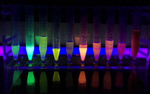
Can these limitations be overcome? Yes, they can and the key to unlock this door was the development of biosensor technology based on genetically-encoded fluorescent proteins (FP) [6, 7]. FP are engineered into thiol-redox biosensors by introducing a couple of vicinal cysteine residues at the protein surface and in close proximity to residues interacting with the hidden chromophore [8]. The cysteines are the biological recognition element that will “sense” and equilibrate their thiol-disulfide state with that of the surrounding environment. The modification of the redox state of these cysteines is accompanied by small and local conformational changes that modify the protonation state of the chromophore and, hence, its spectral properties [6].

A Danish group headed by Jakob R. Winther pioneered the generation and use of the first thiol-redox yellow fluorescent protein biosensor that proved useful in informing the thiol-redox status of Escherichia coli and yeast [9, 10]. Since then, a manifold of articles reported the generation of new redox sensitive FP variants that not only cover almost the full visible spectra (400-700 nm) but also were fine-tuned to operate in organelles with different redox potentials [5, 6, 7].

Depending on their photophysical properties, the FP-redox biosensors can be classified into intensiometric (single wavelength indicators that exhibit changes in their fluorescence intensity) or ratiometric (dual wavelength indicators that exhibit a shift in either their optimum absorption or emission wavelength intensities). The first are suitable for qualitative measurements because the fluorescence signal will depend not only on the concentration of the target molecule but on the expression level of the biosensor and other factors such as cell thickness and pH. The ratiometric biosensors are preferred for quantitative measurements because the interferences pointed above are cancelled out.

A major achievement in this area have been the development of biosensors coupled to different thiol-disulfide oxidoreductases (glutaredoxin, thioredoxin, mycoredoxin and bacilliredoxin) and peroxidases. Fusion to these redox active enzymes expanded the specificity for different redox species (glutathione, mycothiol, bacillithiol, proteins, and peroxides) and, due to kinetic acceleration, lowered the time scale at which redox events can be measured [e.g. from minutes to (nano)seconds].

Because they are genetically encoded, the biosensors can be expressed transiently (as episome) or stably (inducible or constitutively), the last upon integration of the reporter gene into the genome of the target cell/organism. The generation of stable cell lines allows selecting cell populations with a known and homogeneous expression level of the biosensor. At first glance, the establishment of stable redox-reporter cell lines may appear time consuming. However, it constitutes an unlimited source of material that guarantee more consistent results by bypassing the cytotoxic and epigenetic effects, and the cell-to-cell variability associated to uncontrolled transient expression-based assays.

Fluorimeter (plate reader), fluorescent microscope and flow cytometer are suited instruments to detect signals from FP biosensors. The first may be less sensitive and, hence, require a stronger expression of the reporter gene. Confocal microscopy allows for high spatiotemporal resolution at (sub)cellular level, whereas flow cytometry offers statistical robustness and high-throughput power.

With the exception of Archaea, the redox biosensors were expressed in a wide range of organisms from different Kingdoms and, so far, proved valuable to investigate fundamental questions of redox biology [6].

A bright future. The FP-based redox biosensors feature several advantages summarized in: they are produced by the cell and become functional without further intervention by the researcher and can be targeted to almost any subcellular domain/compartment wherefrom they will inform the thiol-redox state in a non-invasive, real-time and dynamic fashion. As long as there is not spectral overlap, the redox biosensors are compatible with multiparametric analysis relying on the use of complementary fluorescent probes or methods. The possibility of tagging the biosensor to different proteins opens the opportunity to “spy” redox signaling processes that control distinct cellular functions with high molecular and temporal precision. The FP biosensors can be rated as excellent biotools to address major questions in the field of redox biochemistry and biology for many pathophysiological phenomena of interest in transmissible (e.g. infectious) and non-transmissible disease’s models. Last but not least, they also show great potential in the drug discovery area, with applications that encompass the screening of drug libraries (e.g. for the selection or discard of molecules causing redox unbalance) and the study of drug toxicity [11] and mode of action [12].


References

  1. G. Salinas, M. A. Comini. Alternative Thiol-Based Redox Systems Antioxidants & Redox Signaling, 28(6): 407–9, 2018. | doi: 10.1089/ars.2017.7464
  2. C. Klomsiri, P. A. Karplus, L. B. Poole. Cysteine-Based Redox Switches in Enzymes Antioxidants & Redox Signaling, 14(6): 1065–77, 2011. | doi: 10.1089/ars.2010.3376
  3. Y. Go, D. P. Jones. Redox compartmentalization in eukaryotic cells Biochimica et Biophysica Acta (BBA) - General Subjects, 1780(11): 1273–90, 2008. | doi: 10.1016/j.bbagen.2008.01.011
  4. M. A. Comini. Measurement and meaning of cellular thiol:disufhide redox status Free Radical Research, 50(2): 246–71, 2016. | doi: 10.3109/10715762.2015.1110241
  5. A. J. Meyer, T. P. Dick. Fluorescent Protein-Based Redox Probes Antioxidants & Redox Signaling, 13(5): 621–50, 2010. | doi: 10.1089/ars.2009.2948
  6. M. Schwarzländer, T. P. Dick, A. J. Meyer, B. Morgan. Dissecting Redox Biology Using Fluorescent Protein Sensors Antioxidants & Redox Signaling, 24(13): 680–712, 2016. | doi: 10.1089/ars.2015.6266
  7. C. V. Piattoni, F. Sardi, F. Klein, S. Pantano, M. Bollati-Fogolin, M. Comini. New red-shifted fluorescent biosensor for monitoring intracellular redox changes Free Radical Biology and Medicine, 134: 545–54, 2019. | doi: 10.1016/j.freeradbiomed.2019.01.035
  8. H. Ostergaard. Shedding light on disulfide bond formation: engineering a redox switch in green fluorescent protein The EMBO Journal, 20(21): 5853–62, 2001. | doi: 10.1093/emboj/20.21.5853
  9. H. Østergaard, C. Tachibana, J. R. Winther. Monitoring disulfide bond formation in the eukaryotic cytosol The Journal of Cell Biology, 166(3): 337–45, 2004. | doi: 10.1083/jcb.200402120
  10. R. Wittig, V. Richter, S. Wittig-Blaich, P. Weber, W. S. L. Strauss, T. Bruns, T. P. Dick, H. Schneckenburger. Biosensor-Expressing Spheroid Cultures for Imaging of Drug-Induced Effects in Three Dimensions Journal of Biomolecular Screening, 18(6): 736–43, 2013. | doi: 10.1177/1087057113480525
  11. J. Franco, F. Sardi, L. Szilágyi, K. E. Kövér, K. Fehér, M. A. Comini. Diglycosyl diselenides alter redox homeostasis and glucose consumption of infective African trypanosomes International Journal for Parasitology: Drugs and Drug Resistance, 7(3): 303–13, 2017. | doi: 10.1016/j.ijpddr.2017.08.001

Marcelo Comini (mcomini@pasteur.edu.uy) holds a degree in Biochemistry (1999) from the Universidad Nacional del Litoral (Santa Fe, Argentina) and a Dr. rerum naturarum (2004) from the Technical University of Braunschweig (Braunschweig, Germany), with a postdoctoral period (2004-2007) at the Biochemistry Centre from the Heidelberg University (Heidelberg, Germany). In 2008, he joined the Institut Pasteur de Montevideo as “Young group leader” of the Laboratory Redox Biology of Trypanosomes and five years later became Principal Investigator. He is a foundational member of the Molecular, Cellular and Animal Technology Program (ProTeMCA) from IP-Montevideo. He was invited to join several European and International consortia devoted to drug discovery research against neglected diseases. He is member of the Sistema Nacional de Investigadores (level 2) and Programa de Desarrollo de las Ciencias Básicas (level 4) from Uruguay. His scientific contributions (59 research articles and 9 book chapters) were published in top peer-reviewed journals and books. The research interest of his team embraces: understanding fundamental aspects of the redox biology of trypanosomatids, the early phase of drug discovery against these pathogens and the development of fluorescent protein-based biosensors.

Add new comment

Demolishing is critical for rebuilding: Is the free 20S proteasome able to degrade proteins in the absence of its regulatory units in vivo?

Submitted by redoxoma on Thu, 02/28/2019 - 15:28
The proteasome 20S

Redoxoma Highlights by Marilene Demasi and Fernanda M. Cunha

Protein degradation is a vital cellular function, needed to enable cells to get rid of misfolded and toxic proteins, to dispose of unused protein components, to control cell cycle and inflammation or simply to demolish the existing cell milieu for metabolic needs or during cell/tissue remodeling. the proteasome (…) is a crucial regulator of cellular protein degradation and homeostasis A major protein complex, the proteasome, composed of several subunits, is a crucial regulator of cellular protein degradation and homeostasis. How the proteasome is regulated and whether it specifically degrades redox-modified proteins is a matter of debate. These mechanisms were put into a critical perspective in a recent review article by Redoxoma investigators (Demasi & da Cunha, 2018).

The ubiquitin-proteasome system is the main pathway of intracellular proteins degradation. It requires the conjugation of poly-ubiquitin chains to target proteins, followed by their recognition by the 19S proteasome regulatory unit coupled to the 20S catalytic core particle. Intracellularly, the 20S proteasome stays coupled to its 19S regulatory subunit, which is the most abundant, in addition to the other regulatory units 11S and PA200. ATP is required in the context of protein ubiquitylation, unfolding and translocation into the 20S catalytic chamber, since all such processes are energy-consuming. In addition to the complete assembled particle, a free pool of the 20S catalytic unit has been proposed to occur intracellularly, but its demonstration has been challenging. Existence of this pool was suggested in the 1980's and demonstrated in vitro. By then, it was shown that free 20S catalytic core would be responsible for the degradation of oxidized proteins, thus independently of the 19S regulatory unit and, consequently, of poly-ubiquitylation and ATP. Since then, a number of independent investigators have reported that not only oxidized but also intrinsically disordered proteins can be degraded by free 20S proteasome in vitro.

Despite those evidences, the hypothesis of protein degradation by a free pool of the 20S proteasome in vivo has remained controversial in the literature. protein degradation by a free pool of the 20S proteasome in vivo has remained controversial in the literature We critically reviewed several experimental data that support such hypothesis. From one side of this question, it is clear that participation of the proteasome in cellular proteolysis goes beyond the classical model of protein poly-ubiquitylation. Indeed, protein degradation through the proteasome may proceed through multiple proteasome complexes, with different requirements, a fact that is sometimes overlooked by part of the scientific community. In the case of the free 20S proteasome, its gate opening is a mandatory and crucial event allowing substrate degradation, but there is not much data on what regulates this process. Our analysis concluded that despite the evidences, few contributions produced unequivocal data supporting in vivo proteolysis through the free 20S proteasome, rendering this issue a meritorious focus of further investigation.


Related article:

  • M. Demasi, F. M. da Cunha. The physiological role of the free 20S proteasome in protein degradation: A critical review Biochimica et Biophysica Acta (BBA) - General Subjects, 1862(12): 2948–54, 2018 | doi: 10.1016/j.bbagen.2018.09.009

Marilene Demasi (marim-I-am-here-asi@b@hotmail.comutantan.gov.br),
Ph.D. Professor at Laboratory of Biochemistry and Biophysics,
Instituto Butantan, Brazil
and
Fernanda M. da Cunha (phernandac-I-am-here-unha@hotmail.com@gmail.com),
Ph.D. Professor at Department of Biochemistry,
Paulista School of Medicine, Federal University of São Paulo, Brazil


Add new comment

A molecular pathway for Protein disulfide isomerase effects on vascular Nox1-dependent oxidant generation

Submitted by redoxoma on Thu, 02/28/2019 - 14:43
Cardiovascular disease

Redoxoma Highlights by Lucia R. Lopes

Superoxide and hydrogen peroxide are important signaling intermediates in cell physiology and local imbalances in their production/removal rates promote oxidative stress, which is a major contributing factor for cardiovascular disease. The primary enzymatic sources of superoxide/hydrogen peroxide with signaling purposes in vascular cells are the Nox (NADPH oxidase) family complexes. Nox1 isoform constitutes a catalytic core at the cell membrane and is constitutively activated in cells (e.g. vascular cells) upon the expression of its canonical regulatory subunits NoxO1 (organizing subunit 1) and NoxA1 (Nox activating subunit 1) [1]. In contrast, p47phox and p67phox homologous to NoxO1 and NoxA1, respectively allow for inducible activation of Nox1 [2]. This is dependent on the phosphorylation of p47phox which produces its conformational change allowing its binding to p22phox and p67phox , culminating in Nox1 activation. Understanding mechanisms whereby Nox catalytic and regulatory subunits are assembled and their expression modulated in physiological or pathological scenarios is a major goal of redox biomedicine.

Protein disulfide isomerase (PDI) belongs to the thioredoxin superfamily of dithiol-disulfide oxidoreductases and its canonical function is to catalyze the correct insertion of disulfide bonds in nascent proteins at the endoplasmic reticulum. Previous work developed at our Redoxoma groups have shown that PDI exerts a critical role in Nox regulation, being essential for growth factor-dependent activation and expression of Nox1, as well as for Nox1-dependent vascular cell migration [3]. However, the mechanisms whereby PDI regulate Nox1 have remained unclear. In a collaborative study with Prof. Francis Miller from Duke University [4], we have now provided a compelling molecular mechanism for PDI-mediated Nox1 regulation. In a series of experiments, we showed that the Nox subunit p47phox is a relevant target of PDI effects. In vitro experiments with recombinant enzymes and mass spectrometry techniques showed that PDI physically interacts with p47phox, forming cross-linked disulfides, an interaction that facilitates p47phox phosphorylation and ensuing Nox1 activation. Cysteine 196 in p47phox and cysteine 400 at the redox active site of PDI are essential for the activation of Nox1 by PDI. Transfection of vascular smooth muscle cells (VSMC) with those proteins mutated in such critical cysteine residues prevented Nox1 activation and VSMC migration in response to the growth factor/vasoconstrictor agonist angiotensin II. Such effects were similar to those described for inhibition/silencing of PDI. Further experiments indicated that mechanisms whereby PDI enhance p47phox phosphorylation involve a reduction of an intramolecular disulfide bond between cys196 and 378 in p47phox, allowing further phosphorylation of the protein and increasing the activation of Nox1. Our findings of increased PDI and Nox1 expression in atherosclerotic aortae and of PDI interaction with p47phox in injured arteries suggest that PDI interaction with p47phox is a relevant mechanism for the effects of PDI as a redox activator of Nox1 in the vasculature. Accordingly, we found a positive correlation between the expression of PDI and p47phox in human atheroma plaques. Altogether, these in vivo data indicate that PDI interaction with p47phox contributes to oxidant generation in contexts related to atherosclerosis 4. Such a novel interaction may provide potential therapeutic targets against vascular disease.

Normal -> Atherosclerosis

References

  1. B. Bánfi, R. A. Clark, K. Steger, K. Krause. Two Novel Proteins Activate Superoxide Generation by the NADPH Oxidase NOX1 Journal of Biological Chemistry, 278(6): 3510–3, 2002 | doi: 10.1074/jbc.c200613200
  2. M. Geiszt, K. Lekstrom, J. Witta, T. L. Leto. Proteins Homologous to p47phoxand p67phoxSupport Superoxide Production by NAD(P)H Oxidase 1 in Colon Epithelial Cells Journal of Biological Chemistry, 278(22): 20006–12, 2003 | doi: 10.1074/jbc.m301289200
  3. L. A. Pescatore, D. Bonatto, F. L. Forti, A. Sadok, H. Kovacic, F. R. M. Laurindo. Protein Disulfide Isomerase Is Required for Platelet-derived Growth Factor-induced Vascular Smooth Muscle Cell Migration, Nox1 NADPH Oxidase Expression, and RhoGTPase Activation Journal of Biological Chemistry, 287(35): 29290–300, 2012 | doi: 10.1074/jbc.m112.394551
  4. M. Gimenez, S. Veríssimo-Filho, I. Wittig, B. M. Schickling, F. Hahner, C. Schürmann, L. E. Netto, J. C. Rosa, R. P. Brandes, S. Sartoretto, et al. Redox Activation of Nox1 (NADPH Oxidase 1) Involves an Intermolecular Disulfide Bond Between Protein Disulfide Isomerase and p47 Arteriosclerosis, Thrombosis, and Vascular Biology, 39(2): 224–36, 2019 | doi: 10.1161/atvbaha.118.311038

Lucia Rossetti Lopes (llo-I-am-here-pes@u@gmail.comsp.br),
Associate Professor at Department of Pharmacology,
Instituto de Ciências Biomédicas, University of São Paulo, Brazil


Cover article image by Scientific Animations licensed under Creative Commons “BY-SA (Attribution-ShareAlike 4.0 International)”

Add new comment

Protein aggregation by aldehydes derived from cholesterol oxidation

Submitted by redoxoma on Tue, 12/11/2018 - 11:15
Protein aggregation by aldehydes derived from cholesterol oxidation

Redoxoma Highlights by Sayuri Miyamoto

Cholesterol is present at a high concentration in the central nervous system. Our brain produces most of its own cholesterol since blood lipoproteins cannot cross the blood-brain barrier. Being one of the major lipids present in myelin, cholesterol is critical for adequate transmission of neural electrical signals and proper neuronal function. A number of studies have shown alterations in cholesterol levels in neurodegenerative diseases. Yet mechanistic aspects connecting cholesterol with neurodegenerative diseases are yet incompletely elucidated.studies have shown alterations in cholesterol levels in neurodegenerative diseases

A hallmark of neurodegenerative diseases is the accumulation of toxic protein clumps or aggregates and increased features of oxidative stress. Amyotrophic lateral sclerosis (ALS) is a complex neurodegenerative disease in which neurons from both spinal cord and brain are affected. To date pathological mechanisms leading to progressive motor neuron degeneration still remains to be clarified.

In order to get some new insights into ALS disease mechanism, we investigated whether cholesterol oxidation could have any connection with this devastating disease. Cholesterol can be oxidized by enzymatic and non-enzymatic mechanisms to generate oxidized derivatives collectively known as oxysterols. An interesting feature of cholesterol oxidation by free radicals and particularly by singlet oxygen is the production of reactive products bearing an aldehyde group (secosterol A and B), which are highly prone to bind to proteins.

Using ALS rat model having SOD1 mutation, we first investigated the presence of cholesterol aldehydes in brain, spinal cord and blood plasma of these animals. Interestingly, cholesterol aldehydes were ubiquitously present in all analyzed tissues. Of note, increased aldehyde levels were detected in the blood plasma of symptomatic animals compared to non-symptomatic ones, suggesting that cholesterol oxidation might be exacerbated during disease progression.

cholesterol aldehydes could irreversibly modify SOD1 proteins, enhancing their propensity to aggregateThese results stimulated us to conduct a series of in vitro studies to deeply characterize cholesterol aldehyde's effect on protein aggregation. Using SOD1 as a model, it was possible to demonstrate that aldehydes accelerate SOD1 aggregation producing large amorphous aggregates. Furthermore, extensive mass spectrometry analysis of the modified protein revealed that aldehydes bind to specific lysine residues located mostly at the electrostatic loop as well as some residues at the protein dimer interface. Collectively, these results reinforced our hypothesis that cholesterol aldehydes could irreversibly modify SOD1 proteins, enhancing their propensity to aggregate during the course of the disease. Now we are further exploring the potential role of cholesterol aldehyde-induced protein aggregation in vivo.


Related article:

  • L. S. Dantas, A. B. Chaves-Filho, F. R. Coelho, T. C. Genaro-Mattos, K. A. Tallman, N. A. Porter, O. Augusto, S. Miyamoto. Cholesterol secosterol aldehyde adduction and aggregation of Cu,Zn-superoxide dismutase: Potential implications in ALS Redox Biology, 19: 105–15, 2018 | doi: 10.1016/j.redox.2018.08.007

Sayuri Miyamoto, Ph.D. Assistant Professor at Department of Biochemistry,
Institute of Chemistry, University of São Paulo, Brazil


Add new comment