PDI

To adhere or not to adhere? This is the disulfide exchange question!

Submitted by redoxoma on
Foto by Chris Reading (https://pixabay.com/users/chrisreadingfoto-2723427/), under Pixabay License

Highlights by Marcela Franco Mineiro, PhD, from Instituto de Química da USP

Marcela got her PhD in 2019 under supervision of Flavia Carla Meotti at the Laboratory of Redox Processes in Inflammation, Department of Biochemistry, Institute of Chemistry, University of São Paulo, Brazil

The term ‘adhesion’ is simply defined as ‘steady or firm attachment’ [1]. In cell biology, however, the adhesion process is far more complex and involves countless well-orchestrated reactions and interactions between proteins at both extra and intracellular surface of the plasma membrane. Intra and intermolecular disulfide exchange in these proteins is one of the most relevant mechanism that drives cell adhesion/detachment. Therefore, oxidizing and reducing agents can directly affect cell adhesion. Nonetheless, it is hard to infer whether an oxidant would increase or decrease this phenomenon because adhesion involves disulfide linkage/break at specific cysteine residues within each protein. It is well known that catalytic cysteines from extracellular cell surface protein disulfide isomerases (PDIs, of which the prototype is PDIA1, refered to as PDI) are crucial in cell adhesion and disulfide exchange between PDI and integrin takes place during the adhesion process [2, 3]. Of relevance, our group has shown that cysteines at the catalytic site of PDI are rapidly oxidized by urate hydroperoxide (6 × 10³ M⁻¹s⁻¹) [4]; this is a peroxide generated from uric acid in the inflammatory oxidative burst [5]. In fact, the oxidation of PDI by urate hydroperoxide is much faster than that by glutathione disulfide (188 M⁻¹s⁻¹) [6] or hydrogen peroxide (17.3 M⁻¹s⁻¹) [7], but slower than the oxidation by peroxynitrite (6.9 × 10⁴ M⁻¹s⁻¹) [7]. We found that the oxidation of extracellular cell surface PDI by urate hydroperoxide impaired the adherence of vascular endothelial cells to fibronectin in the same way as the thiol alkylating p-CMBS, the PDI inhibitor Rutin and the integrin blocking peptide RGDS. Interestingly, adhesion was markedly inhibited in the first 30 min and, to the exception of the treatment with the irreversibly thiol alkylating p-CMBS, cells were able to adhere after 90 min of treatment [4]. These results show that the oxidation of thiols and inhibition of PDI or integrin disrupt cellular adhesion in a transient way. However, the continuous production of oxidants, as in vascular inflammation, might further recover cell adhesion. Since urate hydroperoxide can be formed extracellularly and efficiently targets cell surface PDI affecting cell adhesion, it might be a mechanism underlying the known vascular endothelial dysfunction described for uric acid. Analogous effects of other vascular oxidants in cell adhesion remain to be investigated.


References

  1. Merriam-Webster [on-line] 2020.url: https://www.merriam-webster.com/dictionary/adhesion
  2. N. Rosenberg, R. Mor-Cohen, V. H. Sheptovitsky, O. Romanenco, O. Hess, J. Lahav. Integrin-mediated cell adhesion requires extracellular disulfide exchange regulated by protein disulfide isomerase Experimental Cell Research, 381(1): 77–85, 2019. | doi: 10.1016/j.yexcr.2019.04.017
  3. A. I. Soares Moretti, F. R. Martins Laurindo. Protein disulfide isomerases: Redox connections in and out of the endoplasmic reticulum Archives of Biochemistry and Biophysics, 617: 106–19, 2017. | doi: 10.1016/j.abb.2016.11.007
  4. M. F. Mineiro, E. de S. Patricio, Á. S. Peixoto, T. L. S. Araujo, R. P. da Silva, A. I. S. Moretti, F. S. Lima, F. R. M. Laurindo, F. C. Meotti. Urate hydroperoxide oxidizes endothelial cell surface protein disulfide isomerase-A1 and impairs adherence Biochimica et Biophysica Acta (BBA) - General Subjects, 1864(3): 129481, 2020. | doi: 10.1016/j.bbagen.2019.129481
  5. R. P. Silva, L. A. Carvalho, E. S. Patricio, J. P. Bonifacio, A. B. Chaves-Filho, S. Miyamoto, F. C. Meotti. Identification of urate hydroperoxide in neutrophils: A novel pro-oxidant generated in inflammatory conditions Free Radical Biology and Medicine, 126: 177–86, 2018. | doi: 10.1016/j.freeradbiomed.2018.08.011
  6. Á. S. Peixoto, R. R. Geyer, A. Iqbal, D. R. Truzzi, A. I. Soares Moretti, F. R. M. Laurindo, O. Augusto. Peroxynitrite preferentially oxidizes the dithiol redox motifs of protein-disulfide isomerase Journal of Biological Chemistry, 293(4): 1450–65, 2017. | doi: 10.1074/jbc.m117.807016
  7. A. Lappi, L. W. Ruddock. Reexamination of the Role of Interplay between Glutathione and Protein Disulfide Isomerase Journal of Molecular Biology, 409(2): 238–49, 2011. | doi: 10.1016/j.jmb.2011.03.024
Add new comment

A molecular pathway for Protein disulfide isomerase effects on vascular Nox1-dependent oxidant generation

Submitted by redoxoma on
Cardiovascular disease

Redoxoma Highlights by Lucia R. Lopes

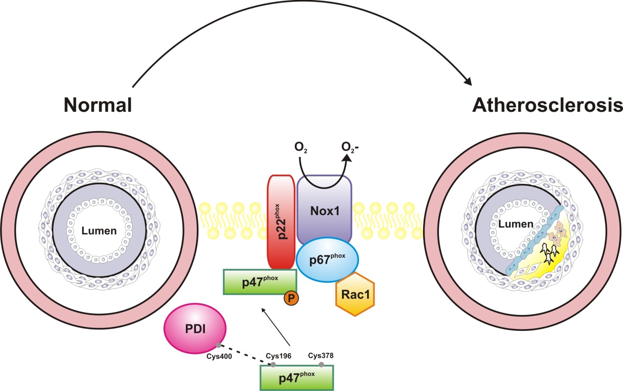
Superoxide and hydrogen peroxide are important signaling intermediates in cell physiology and local imbalances in their production/removal rates promote oxidative stress, which is a major contributing factor for cardiovascular disease. The primary enzymatic sources of superoxide/hydrogen peroxide with signaling purposes in vascular cells are the Nox (NADPH oxidase) family complexes. Nox1 isoform constitutes a catalytic core at the cell membrane and is constitutively activated in cells (e.g. vascular cells) upon the expression of its canonical regulatory subunits NoxO1 (organizing subunit 1) and NoxA1 (Nox activating subunit 1) [1]. In contrast, p47phox and p67phox homologous to NoxO1 and NoxA1, respectively allow for inducible activation of Nox1 [2]. This is dependent on the phosphorylation of p47phox which produces its conformational change allowing its binding to p22phox and p67phox , culminating in Nox1 activation. Understanding mechanisms whereby Nox catalytic and regulatory subunits are assembled and their expression modulated in physiological or pathological scenarios is a major goal of redox biomedicine.

Protein disulfide isomerase (PDI) belongs to the thioredoxin superfamily of dithiol-disulfide oxidoreductases and its canonical function is to catalyze the correct insertion of disulfide bonds in nascent proteins at the endoplasmic reticulum. Previous work developed at our Redoxoma groups have shown that PDI exerts a critical role in Nox regulation, being essential for growth factor-dependent activation and expression of Nox1, as well as for Nox1-dependent vascular cell migration [3]. However, the mechanisms whereby PDI regulate Nox1 have remained unclear. In a collaborative study with Prof. Francis Miller from Duke University [4], we have now provided a compelling molecular mechanism for PDI-mediated Nox1 regulation. In a series of experiments, we showed that the Nox subunit p47phox is a relevant target of PDI effects. In vitro experiments with recombinant enzymes and mass spectrometry techniques showed that PDI physically interacts with p47phox, forming cross-linked disulfides, an interaction that facilitates p47phox phosphorylation and ensuing Nox1 activation. Cysteine 196 in p47phox and cysteine 400 at the redox active site of PDI are essential for the activation of Nox1 by PDI. Transfection of vascular smooth muscle cells (VSMC) with those proteins mutated in such critical cysteine residues prevented Nox1 activation and VSMC migration in response to the growth factor/vasoconstrictor agonist angiotensin II. Such effects were similar to those described for inhibition/silencing of PDI. Further experiments indicated that mechanisms whereby PDI enhance p47phox phosphorylation involve a reduction of an intramolecular disulfide bond between cys196 and 378 in p47phox, allowing further phosphorylation of the protein and increasing the activation of Nox1. Our findings of increased PDI and Nox1 expression in atherosclerotic aortae and of PDI interaction with p47phox in injured arteries suggest that PDI interaction with p47phox is a relevant mechanism for the effects of PDI as a redox activator of Nox1 in the vasculature. Accordingly, we found a positive correlation between the expression of PDI and p47phox in human atheroma plaques. Altogether, these in vivo data indicate that PDI interaction with p47phox contributes to oxidant generation in contexts related to atherosclerosis 4. Such a novel interaction may provide potential therapeutic targets against vascular disease.

Normal -> Atherosclerosis

References

  1. B. Bánfi, R. A. Clark, K. Steger, K. Krause. Two Novel Proteins Activate Superoxide Generation by the NADPH Oxidase NOX1 Journal of Biological Chemistry, 278(6): 3510–3, 2002 | doi: 10.1074/jbc.c200613200
  2. M. Geiszt, K. Lekstrom, J. Witta, T. L. Leto. Proteins Homologous to p47phoxand p67phoxSupport Superoxide Production by NAD(P)H Oxidase 1 in Colon Epithelial Cells Journal of Biological Chemistry, 278(22): 20006–12, 2003 | doi: 10.1074/jbc.m301289200
  3. L. A. Pescatore, D. Bonatto, F. L. Forti, A. Sadok, H. Kovacic, F. R. M. Laurindo. Protein Disulfide Isomerase Is Required for Platelet-derived Growth Factor-induced Vascular Smooth Muscle Cell Migration, Nox1 NADPH Oxidase Expression, and RhoGTPase Activation Journal of Biological Chemistry, 287(35): 29290–300, 2012 | doi: 10.1074/jbc.m112.394551
  4. M. Gimenez, S. Veríssimo-Filho, I. Wittig, B. M. Schickling, F. Hahner, C. Schürmann, L. E. Netto, J. C. Rosa, R. P. Brandes, S. Sartoretto, et al. Redox Activation of Nox1 (NADPH Oxidase 1) Involves an Intermolecular Disulfide Bond Between Protein Disulfide Isomerase and p47 Arteriosclerosis, Thrombosis, and Vascular Biology, 39(2): 224–36, 2019 | doi: 10.1161/atvbaha.118.311038

Lucia Rossetti Lopes (llo-I-am-here-pes@u@gmail.comsp.br),
Associate Professor at Department of Pharmacology,
Instituto de Ciências Biomédicas, University of São Paulo, Brazil


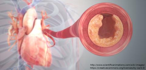
Cover article image by Scientific Animations licensed under Creative Commons “BY-SA (Attribution-ShareAlike 4.0 International)”

Add new comment