
Highlights

Peroxiredoxin hyperoxidation increases in the presence of bicarbonate/carbon dioxide

Submitted by redoxoma on Fri, 02/28/2020 - 18:34
peroxidase cycle, hyperoxidation pathway and interaction and oxidation of other thiol proteins, signaling

Highlights by Daniela R. Truzzi and Ohara Augusto, from Instituto de Química da USP
Corresponding author e-mail: dtruz-I-am-here-zi@hotmail.com@iq.usp.br

Peroxiredoxins (Prx) are abundant thiol peroxidases that react rapidly with H2O2, constituting an important antioxidant defense and acting as sensors and transmitters of H2O2 signals in cells. Eukariotic 2-Cys Prxs lose their peroxidase activity at high hydroperoxide levels in a process called hyperoxidation, which constitutes a pathway to Prx functions beyond the antioxidant activity (Fig. 1) [1]. Since the biologically ubiquitous HCO3/CO2 pair accelerates the reaction of H2O2 with several biothiols [2, 3], we examined whether physiological concentrations of the HCO3/CO2 pair (25 mM) could increase recombinant human peroxiredoxin 1 (Prx1) hyperoxidation by H2O2 [4]. Immunoblotting, kinetic and MS/MS experiments revealed that HCO3/CO2 increases Prx1 hyperoxidation and inactivation both in the presence of excess H2O2 and during enzymatic (NADPH/thioredoxin reductase/thioredoxin) and chemical (dithiothreitol) turnover. Based on previous studies, we hypothesized that the stimulating effect of HCO3/CO2 was due to HCO4 (peroxymonocarbonate), a peroxide present in equilibrated solutions of H2O2 and HCO3/CO2 [2, 3]. Indeed, additional experiments and calculations uncovered that HCO4 oxidizes CPSOH to CPSO2 with a second-order rate constant two orders of magnitude higher than H2O2 ((1.5 ± 0.1) × 10⁵ and (2.9 ± 0.2) × 10³ M⁻¹.s⁻¹, respectively) and that HCO4 is 250 times more efficient than H2O2 at inactivating 1% Prx1 per turnover [4]. The fact that the biologically ubiquitous HCO3/CO2 pair stimulates Prx1 hyperoxidation may be quite relevant to cell homeostasis because the antioxidant and redox relay functions of the enzyme decline but other actions may rise, such as the chaperone-like activity, the redox signaling pathways mediated by Cys-based proteins that are poorly reactive towards H2O2 and the maintenance of Trx-dependent activities (Fig. 1). Relevantly, parallel studies led by Christine Winterbourn and co-workers reported that the HCO3/CO2 pair also stimulates Prx2 and Prx3 hyperoxidation [5] and protein tyrosine phosphatase 1B inactivation involved in epidermal growth factormediated signaling [6]. Taking together, these recent studies confirm that HCO4 deserves further investigation as a biological oxidant [3] and point to a possible role of HCO3/CO2 levels in H2O2mediated signaling.

Peroxidase cycle, hyperoxidation pathway


Figure 1. Simplified scheme of the catalytic cycle and the hyperoxidation pathway of 2-Cys Prxs and the activities related to them.


References

  1. E. A. Veal, Z. E. Underwood, L. E. Tomalin, B. A. Morgan, C. S. Pillay. Hyperoxidation of Peroxiredoxins: Gain or Loss of Function? Antioxidants & Redox Signaling, 28(7): 574–90, 2018. | doi: 10.1089/ars.2017.7214
  2. D. F. Trindade, G. Cerchiaro, O. Augusto. A Role for Peroxymonocarbonate in the Stimulation of Biothiol Peroxidation by the Bicarbonate/Carbon Dioxide Pair Chemical Research in Toxicology, 19(11): 1475–82, 2006. | doi: 10.1021/tx060146x
  3. D. R. Truzzi, O. Augusto. Influence of CO2 on Hydroperoxide Metabolism Hydrogen Peroxide Metabolism in Health and Disease, (Vissers, M.C.M., Hampton, M., Kettle, A.J. eds) 81–99, Oxidative stress and disease, Taylor & Francis/CRC Press, Boca Raton, 2017. | doi: 10.1201/9781315154831-4
  4. D. R. Truzzi, F. R. Coelho, V. Paviani, S. V. Alves, L. E. S. Netto, O. Augusto. The bicarbonate/carbon dioxide pair increases hydrogen peroxide-mediated hyperoxidation of human peroxiredoxin 1 Journal of Biological Chemistry, 294(38): 14055–67, 2019. | doi: 10.1074/jbc.ra119.008825
  5. A. V. Peskin, P. E. Pace, C. C. Winterbourn. Enhanced hyperoxidation of peroxiredoxin 2 and peroxiredoxin 3 in the presence of bicarbonate/CO2 Free Radical Biology and Medicine, 145: 1–7, 2019. | doi: 10.1016/j.freeradbiomed.2019.09.010
  6. M. Dagnell, Q. Cheng, S. H. M. Rizvi, P. E. Pace, B. Boivin, C. C. Winterbourn, E. S. J. Arnér. Bicarbonate is essential for protein-tyrosine phosphatase 1B (PTP1B) oxidation and cellular signaling through EGF-triggered phosphorylation cascades Journal of Biological Chemistry, 294(33): 12330–8, 2019. | doi: 10.1074/jbc.ra119.009001

Add new comment

Something looks rotten in the Redox Kingdom… The Emerging Biochemistry of Hydrogen Sulfide and Persulfides

Submitted by redoxoma on Fri, 02/28/2020 - 17:37
Hydrogen sulfide

Main Article by Beatriz Alvarez from Laboratorio de Enzimología, Facultad de Ciencias, Universidad de la República, Montevideo, Uruguay
Correspondig author e-mail: beatri@qualquerz.alvarez@fcien.edu.uy

Highlights. Hydrogen sulfide (H2S/HS) can be formed in mammals and trigger physiological effects. It can react with metal centers and oxidized thiol products such as disulfides (RSSR) and sulfenic acids (RSOH). Reactions with oxidized thiol products form persulfides (RSSH/RSS). Persulfides are more acidic and nucleophilic than thiols and, unlike thiols, they can also be electrophilic. They are also good reductants. The modification of critical cysteines to persulfides has been proposed to transduce the effects of H2S.

Hydrogen sulfide, a molecular relic with novel roles

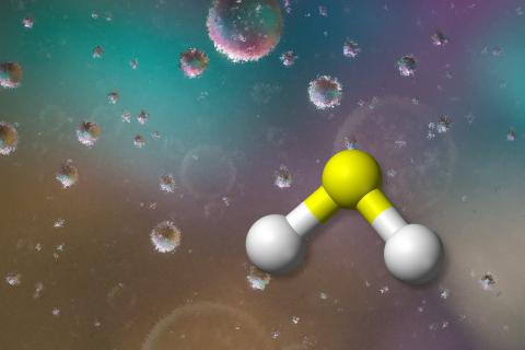
Hydrogen sulfide (H2S) is a gas with the characteristic smell of rotten eggs. Its structure is comparable to water, but due to the lower electronegativity of sulfur with respect to oxygen, H2S is less polar than water and has low intermolecular forces, so it is a gas at room temperature. It is a weak diprotic acid with pKas of 7 and 17. Thus, at physiological pH, 70 % is available as HS and 30 % as H2S.

H2S was present in the environment where life arose, and it was likely part of the first energy-yielding metabolic process. Today, microorganisms can use or form H2S in various ways. For example, lithotrophs use H2S as electron donor to respiratory chains. For several species including mammals, H2S is toxic because it inhibits cellular respiration. This toxicity has been linked to massive extinction events in the history of life on Earth, such as the Permian–Triassic extinction when more than 96 % of marine species were killed. Nevertheless, in the nineties it was recognized that H2S can be synthesized in mammals and that it can exert several physiological effects with potential health benefits. Numerous effects have been reported, including neuromodulation and attenuation of ischemia-reperfusion injury. Yet, the molecular bases of the observed effects are typically unclear.

The formation of H2S in mammals relies on enzymes of sulfur amino acid pathways: cystathionine beta-synthase, cystathionine gamma-lyase and mercaptopyruvate sulfur transferase. Intestinal bacteria can also synthesize it. H2S can be measured in tissues; the reported values have decreased with time, as analytical methods have improved, and are in the tens of nanomolar range. H2S can rapidly traverse membranes. Mitochondria are the main site for H2S catabolism. By the action of several enzymes, H2S fuels electrons to ubiquinone and is oxidized to thiosulfate (S2O32–), sulfite (SO32–) and sulfate (SO42–). Besides, H2S is an inhibitor of cytochrome c oxidase.

H2S (rather, HS) is nucleophilic, which means that it can donate a pair of electrons to form a bond. In this regard, it is analogous to thiols (RSH), although H2S is a slightly weaker nucleophile and the products formed are different. One of the possible reactions of H2S in biological contexts is with metal centers. For example, H2S can bind to ferric heme. The binding can be reversible, as in the case of hemeproteins that transport H2S in invertebrates, or the iron can be reduced by H2S. In some cases, sulfheme is formed, a green variant in which the heme ring is covalently modified. Reactions of H2S with oxidants derived from the partial reduction of oxygen (e.g. H2O2) can occur, but direct scavenging is unlikely to explain antioxidant effects. H2S is also able to react with oxidized derivatives of thiols such as disulfides (RSSR) or sulfenic acids (RSOH). In these reactions, persulfides (RSSH/RSS) are formed (Eqs. 1 and 2).

H2S + RSSR ⇆ RSSH + RSH

Eq. 1

H2S + RSOH → RSSH + H2O

Eq. 2

Importantly, H2S cannot react with reduced thiols. In fact, another reaction that forms persulfides is that of thiols with products of H2S oxidation, such as polysulfides (HSnSH).

Persulfides, possible transducers of the biological effects of H2S

Protein and non-protein persulfides are detected in biological samples and can be formed in thiols through several pathways, some of which are dependent on H2S. The observation that H2S can trigger physiological effects and that it can yield persulfides has led to the proposal that the signaling effects of H2S may in part involve persulfides. Thus, the posttranslational modification of critical protein cysteines to persulfides could unleash downstream effects of H2S. Besides this role in signaling, persulfides participate in several biosynthetic pathways, such as in the assembly of iron-sulfur clusters. In addition, there exist enzymes able to produce (e.g. sulfide quinone oxidoreductase), transfer (e.g. rhodanese) or react (e.g. persulfide dioxygenase) with persulfides.

Persulfides have a very rich biological chemistry that constitutes an open area of investigation (Fig. 1). First, persulfides are more acidic than thiols. This determines that the ionized species (RSS) are highly available at physiological pH. Second, like thiols, persulfides are nucleophilic and can react with electrophiles (E+), although the products that are formed are different, disulfides in the case of persulfides and thioethers in the case of thiols. The ionized species are better nucleophiles than thiolates and have higher availability at physiological pH. Third, unlike thiols, which can only act as nucleophiles, persulfides, when protonated, also have electrophilic character, which can be located in both sulfur atoms. Fourth, they can react with oxidants. They are particularly good one-electron reductants, due in part to the relative stability of the RSS radicals formed. Besides, persulfide formation protects protein thiols from irreversible oxidative damage, since the products of persulfide oxidation (RSSO2H and RSSO3H) can be reduced to thiols.

RSSH <---> RSS- + H+


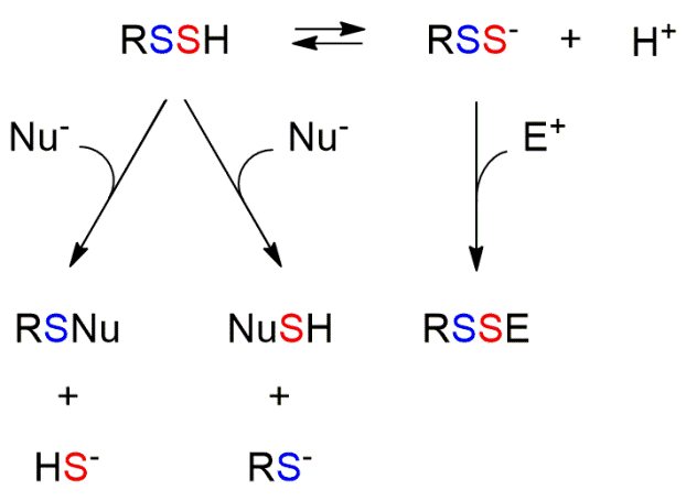
Fig. 1. Reactivity of persulfides

To sum up, persulfides are nowadays raising interest as candidate species for the transduction of the signals triggered by H2S. For further reading on H2S and persulfides, readers are referred to the recent reviews cited below and the references therein. It is likely that the next years will see an increase in the understanding of the biochemistry of these fascinating species.


Further reading

  1. M. R. Filipovic, J. Zivanovic, B. Alvarez, R. Banerjee. Chemical Biology of H2S Signaling through Persulfidation Chemical Reviews, 118(3): 1253–337, 2017. | doi: 10.1021/acs.chemrev.7b00205
  2. D. Benchoam, E. Cuevasanta, M. Möller, B. Alvarez. Hydrogen Sulfide and Persulfides Oxidation by Biologically Relevant Oxidizing Species Antioxidants, 8(2): 48, 2019. | doi: 10.3390/antiox8020048
  3. N. Lau, M. D. Pluth. Reactive sulfur species (RSS): persulfides, polysulfides, potential, and problems Current Opinion in Chemical Biology, 49: 1–8, 2019. | doi: 10.1016/j.cbpa.2018.08.012
  4. D. Benchoam, E. Cuevasanta, M. Möller, B. Alvarez. Persulfides, at the crossroads between hydrogen sulfide and thiols Essays in Biochemistry, 64(1): 155–68, 2020. | doi: 10.1042/ebc20190049
  5. J. M. Fukuto, V. S. Vega, C. Works, J. Lin. The chemical biology of hydrogen sulfide and related hydropersulfides: interactions with biologically relevant metals and metalloproteins Current Opinion in Chemical Biology, 55: 52–8, 2020. | doi: 10.1016/j.cbpa.2019.11.013

Add new comment

Uncovering the antioxidant mechanisms of N-acetyl cysteine

Submitted by redoxoma on Thu, 09/27/2018 - 16:19
N-acetyl-L-cysteine
N-acetyl cysteine is often used as antioxidant, but its mechanism of action is obscure. Novel literature data brings an unexpected turn to this question.

Literature Highlights by Flavia C. Meotti

Investigating the roles of reactive oxygen species in experimental and clinical studies is usually a challenge either to experts in the field or to researchers from other fields. Since these species are, as their name suggest, reactive, they are transient and rarely reach a steady state. Likewise, their track is hard to follow because cells have evolved to rapidly regenerate the intracellular redox state. Therefore, researchers usually opt to use antioxidants to confirm the participation of reactive oxygen species in specific events. A widely used antioxidant is N-acetyl cysteine (NAC), however the exact antioxidant mechanisms of NAC are incompletely known. The traditionally accepted mechanism is that NAC behaves as a source of cysteine to glutathione synthesis. However, the limited increase in GSH levels documented after NAC treatment indicates that NAC may act by alternative pathways to protect against oxidants. Another claimed mechanism is that NAC is a direct scavenger of reactive oxygen species. However, the latter is highly unlikely, because the very low rate constants of these reactions pose a crucial kinetic constraint. Recenly, Ezerina et al provided an elegant paradigm swithc in this subject. NAC was demonstrated to be a source of hydrogen sulfide and to generate hydropersulfides in thiol proteins in the mitochondrial matrix. The authors delineated the intracellular catabolism of NAC and the formation of sulfane sulfur species. These hydropersulfides are more reactive than their respective thiol precursors and can therefore directly scavenger oxidants. A relevant conclusion from this study was that the metabolism of NAC inside cells and production of sulfane sulfur species are likely the main contributors to the rapid and frequently observed antioxidative and cytoprotective effects of NAC. Different thiol proteins will present varied susceptibility to the formation of these hydropersulfides and so NAC might distinctively affect such redox pathways. This certainly should be considered when inferring the effects of NAC as a global antioxidant. If we already knew that reactive intermediate scavenging was not a mechanism of NAC effects but did not have a clear mechanism to cite, now we are well grounded to start considering such a novel mechanism.


Related article:

  • D. Ezeriņa, Y. Takano, K. Hanaoka, Y. Urano, T. P. Dick. N-Acetyl Cysteine Functions as a Fast-Acting Antioxidant by Triggering Intracellular H2S and Sulfane Sulfur Production Cell Chemical Biology, 25(4): 447–59, 2018 | doi: 10.1016/j.chembiol.2018.01.011

Flavia C. Meotti, PhD. Professor at Department of Biochemistry,
Institute of Chemistry, University of São Paulo, Brazil


Add new comment

Is redox metabolism connected with Circadian Rhythm?

Submitted by redoxoma on Tue, 11/22/2016 - 11:47

The Radical-Free Corner by Carolina G. Fernandes

Circadian Rhythm is any biological process that temporally organizes behavioral, physiological, and molecular events around the 24h day-night cycle. This process has evolved over approximately 2.5 billion years ago at the Great Oxidation Event. The later involved increases in atmospheric oxygen levels that simulatenwously enabled organisms to resonate with their environment such that their internal cycles anticipate and match external rhythms on Earth. In mammals, this rhythmicity is controlled by the hypothalamic suprachiasmatic nuclei (SCN) receiving retina signals and “translating” them into each tissue/organ that commonly responds using the transcription/translation feedback loop (TTFL) mechanisms (activating or inhibiting gene transcription). However, recent investigations demonstrated that TTFL pathways are not the only mechanisms to coordinate circadian clocks and point to a role for redox processes in these effects. Specifically, Many of those mechanisms are linked to TTFL pathways and include NAD+ oscillations, which control the rate-limiting enzyme in the NAD+ salvage pathway via feedback mechanisms. In addition, other energy/redox metabolites also oscillate in mammals in a circadian fashion, such as glutathione, NAD(P)H and ATP, also through TTFL mechanisms. However, peroxiredoxins (PRX) also oscillate in circadian rhythm in a highly conserved fashion, since Archea to humans, but even in enucleated cells. Furthermore, peroxiredoxin oscillations persist even in red blood cells, reinforcing the existence of non-TTFL circadian regulation mechanism. The importance of redox circuits in circadian clock regulation fits well with the fact that disturbance in these rhythms is linked to pathological processes such as aging, neurodegenerative disorders, cancer and various metabolic conditions [1], in which redox processes have been proposed to play a significant role.


  1. N. B. Milev and A. B. Reddy. Circadian redox oscillations and metabolism. Trends in Endocrinology & Metabolism, 26: 430-7, 2015 | doi:10.1016/j.tem.2015.05.012

Contributed by Carolina Gonçalves Fernandes, post-doc at the Francisco Laurindo Lab
Heart Institute, University of São Paulo School of Medicine

Tags

Add new comment

The two faces of peroxiredoxins

Submitted by redoxoma on Sat, 10/31/2015 - 20:05

Highlights by Larissa A. C. Carvalho

In the antiquity, many cities protected their territories by surrounding fortifications, with giant gates and arches working as unique gateways. The Roman god Janus, known as the protector of entrances or new-beginnings, is represented by a head with two faces in opposite directions. The two faces of Janus symbolize the duality of nature. Well, nature itself is marked by dichotomy. What in nature cannot be good and bad at the same time? That is the origin of the term Janus effect.

A remarkable example of the Janus effect is the mustard gas (1,1-thiobis-2-chloroethane). Used as a chemical weapon during World War I, it causes serious inflammation of the skin and painful blisters. In high-exposition levels, it acutely causes blindness and destroys the lungs’ alveoli, killing the exposed victim. But, what a coincidence: during World War II, the mustard gas showed its therapeutic face. In 1943, the Liberty ship carrying such gas suffered an attack. The survivors were treated by doctor Cornelius Rhoads, who noticed a dramatic drop in the number of white blood cells, attributing it to the mustard gas inhaled by the crew members. The doctor suggested the use of this compound to treat cancer, especially leukemia, where the increase of leukocytes is brutal. Although nowadays other compounds are safer and more effective than mustard gas, this serves as a reminder of how many compounds can be rediscovered after their first reported effects.

It is well established that the role of peroxiredoxins is to eliminate H2O2 by directly discharging the oxidative equivalents to the thioredoxin system. However, for the first time, Sobotta and colleagues [1] described a new role for human peroxiredoxin 2 as a redox signaling pathway. At the same time it plays its role of detoxifying the organism, it relays oxidative equivalents to a transcription factor, STAT3, inactivating it In response to cytokines and growth factors, STAT3 mediates the expression of many prostimulatory genes such as thos related to cell growth and apoptosis, playing a key role in many cellular processes.

In the past, peroxiredoxins have been found to be associated with many signaling complexes, but it always has been assumed that its association is of a non-specific type or that its role is to remove H2O2 and to protect other proteins from oxidation damage. However, depending on location and context, peroxiredoxins may switch between thioredoxin and other proteins, acting as a dual-face protein: playing scavenging and signaling modes. Therefore, the anti- and pro-oxidant pathways (scavenging and signaling) may be inextricably interconnected through thiol peroxidases. Notwithstanding, could the peroxiredoxin be a new example of the Janus effect? Well, like Barry Halliwell and John Gutteridge wrote in their book Free radicals in biology and medicine, the Neil Young’s song and an undeniable truth: The same thing that makes you live, can kill you in the end.


  1. M. C. Sobotta, W. Liou, S. Stocker, D. Talwar, M. Oehler, T. Ruppert, A. N. Scharf, T. P. Dick. Peroxiredoxin-2 and STAT3 form a redox relay for H2O2 signaling Nature Chemical Biology, 11(1): 64-70, 2015 | doi: 10.1038/nchembio.1695

Larissa Anastácio da Costa Carvalho is a PhD Student from the laboratory of Redox Processes in Inflammatory Response, Instituto de Química da USP, coordinated by Flavia C. Meotti

Tags

Add new comment

The controversy about antibiotic lethality and reactive oxygen species

Submitted by redoxoma on Sat, 10/31/2015 - 20:04

Highlights by José F. Silva Neto

Antibiotics are powerful compounds in our battle against bacterial diseases. Despite their miraculous efficacy over decades, nowadays we are faced with the global spreading of antibiotic resistance and the decrease of our antibiotic arsenal. For many years, we learned that antibiotics exert their effect by direct interaction with different primary bacterial targets, causing killing (bactericidal drugs) or growth inhibition (bacteriostatic drugs). In 2007, an influential paper from the Collins laboratory [1] placed reactive oxygen species (ROS) as central players in the mechanism of cell death induced by bactericidal antibiotics. Ever since, the hypothesis that clinically used antibiotics kill bacteria by stimulating the formation of oxidants has raised an intense scientific debate, with many groups generating experimental evidence that either support [1, 2, 3] or contradict [4, 5, 6] this hypothesis. At the heart of this controversy is the choice of assays used by those different groups, which resulted in ambiguous or contradictory data [7, 8].

The initial model of the Collins group proposed that bactericidal antibiotics of distinct classes, regardless of their primary targets, cause cell death by a common mechanism involving production of the reactive oxygen species hydroxyl radical. These conclusions were mainly supported by the use of a redox-sensitive dye (hydroxyphenyl fluorescein, HPF) intended to detect hydroxyl radical accumulation after antibiotic treatment. Accordingly, a hydroxyl radical quencher (thiourea) and an iron chelator (dipyridyl) suppressed the antibiotic-induced fluorescence [1]. Some years later, a series of works using classical genetic and biochemical strategies challenged this model of antibiotic lethality mediated by oxidants [4, 5, 6]. These papers found that: (i) many bactericidal antibiotics are equally effective against bacteria under aerobic and anaerobic conditions; (ii) the role of thiourea and dipyridyl in protecting cells from antibiotic killing is also observed anaerobically; (iii) antibiotic treatment does not create hydrogen peroxide stress or increase the levels of unincorporated iron; (iv) dye probes used to detect hydroxyl radicals could be nonspecifically oxidized. All these data led authors to refute the original ROS hypothesis of antibiotic toxicity [7]. More recently, a new paper from the Collins group addressed experimentally point-by-point most of the challenges to such ROS hypothesis [3], contributing to strengthen the relation between oxidants and antibiotic lethality [8]. Despite the opposite viewpoints, this controversy is functioning to accelerate our understanding of the complex mechanisms by which antibiotics kill bacteria, and inspiring redox-based strategies [2] to increase the efficacy of our available antibiotics.


  1. M. A. Kohanski, D. J. Dwyer, B Hayete, C. A. Lawrence, J. J. Collins. A common mechanism of cellular death induced by bactericidal antibiotics Cell, 130 (5): 797-810, 2007 | doi: 10.1016/j.cell.2007.06.049
  2. M. P. Brynildsen, J. A. Winkler, C. S. Spina, I. C. MacDonald, J. J. Collins. Potentiating antibacterial activity by predictably enhancing endogenous microbial ROS production Nature Biotechnology, 31 (2): 160-65, 2013 | doi: 10.1038/nbt.2458
  3. D. J. Dwyer, P. A. Belenky, J. H. Yang, I. C. MacDonald, J. D. Martell, N. Takahashi, C. T. Chan, M. A. Lobritz, D. Braff, E. G. Schwarz, J. D. Ye, M. Pati, M. Vercruysse, P. S. Ralifo, K. R. Allison, A. S. Khalil, A. Y. Ting, G. C. Walker, J. J. Collins. Antibiotics induce redox-related physiological alterations as part of their lethality Proceedings of the National Academy of Sciences of the United States of America, 111 (20): E2100-9, 2014 | doi: 10.1073/pnas.1401876111
  4. Y. Liu, J. A. Imlay. Cell death from antibiotics without the involvement of reactive oxygen species Science, 339 (6124): 1210-3, 2013 | doi: 10.1126/science.1232751
  5. Keren, Y. Wu, J. Inocencio, L. R. Mulcahy, K. Lewis. Killing by bactericidal antibiotics does not depend on reactive oxygen species Science, 339 (6124): 1213-6, 2013 | doi: 10.1126/science.1232688
  6. B. Ezraty, A. Vergnes, M. Banzhaf, Y. Duverger, A. Huguenot, A. R. Brochado, S. Y. Su, L. Espinosa, L. Loiseau, B. Py, A. Typas, F. Barras. Fe-S cluster biosynthesis controls uptake of aminoglycosides in a ROS-less death pathway Science, 340 (6140): 1583-7,340 (6140): 1583-7, 2013 | doi: 10.1126/science.1238328
  7. J. A. Imlay. Diagnosing oxidative stress in bacteria: not as easy as you might think Current Opinion in MicrobiologyCurrent Opinion in Microbiology, 24: 124-131,24: 124-131, 2015 | doi: 10.1016/j.mib.2015.01.004
  8. X. Zhao, K. Drlica. Reactive oxygen species and the bacterial response to lethal stress Current Opinion in Microbiology, 21: 1-6, 2014 | doi: 10.1016/j.mib.2014.06.008

José Freire da Silva Neto is a Professor of the Depto. de Biologia Celular e Molecular e Bioagentes Patogênicos, Faculdade de Medicina de Ribeirão Preto, University of São Paulo, and a former post-doctoral fellow at the laboratory of Luis Eduardo Soares Netto, a principal investigator of our CEPID-Redoxoma.

Tags

Add new comment

A comprehensive approach to identify redox and non-redox targets of Trx-like proteins

Submitted by redoxoma on Fri, 05/29/2015 - 20:02

Highlights by Lia S. Nakao

Like the old dictum that says “birds of a feather flock together”, understanding the specific partners of a given protein provides an important clue about its function. Thioredoxin 1 (Trx1) is a well-known redox protein that contains a CXXC motif (cysteines residues flanking two aminoacid residues), responsible for its disulfide reductase function. The first (C-terminal) Cys of the motif attacks the disulfide of the target protein, producing a short lived mixed disulfide, which is reduced by the second (N-terminal resolving) Cys, releasing Trx1 and the target, in the oxidized and reduced forms, respectively. If the resolving Cys is replaced by a non-redox residue, such as alanine or serine, the target will be trapped in the mixed disulfide and can be identified [1]. This is the basis of the mechanism-based, trapping mutant method, which has identified several Trx1 targets. A recent work [2] adapted this strategy to identify novel partners of Trx1 and of other CXXC-containing proteins, namely Rdx12, nucleoredoxin 1 (Nrx1) and thioredoxin-like protein 1 (Txnl1) in cell culture model (HEK293T), by immunoaffinity purification and liquid chromatography/mass spectrometry. Some important steps were included to improve the method: (i) cellular overexpression of Trx1 and Trx-like proteins allowed the trapping of the targets in situ, increasing the possibility of trapping physiological partners; (ii) the endogenous expression of the studied proteins was knocked down to minimize the resolving of the mixed disulfide; (iii) the overexpressed proteins carried both a hemagglutinin (HA) tag to facilitate the immunopurification using anti-HA coupled to beads, and a tobacco etch virus (TEV) protease cleavage site to allow the elution of the immunopurified complex in a very mild condition; (iv) the comparison of the MS data of each candidate across the 3 constructs (the CXXC, CXXS and the SXXC, where S represents the serine amino acid), as well as of the control lacking expression construct, allowed the calculation of target enrichment, supporting a detailed and quantitative analyses of the datasets. The method was validated by the confirmation of peroxiredoxins 1, 2, 4 and 5 as Trx1 targets. This strategy led to identification of apoptosis inducing factor 1 (AIF) and Mia40 (a protein involved with the oxidative import of mitochondrial proteins) as novel Trx1 redox targets, and glutathione peroxidase 4 as a novel Rdx12 redox target, as they were significantly enriched in their CXXS mutants. Nrx1 and Txnl1 CXXS mutants, however, showed low enrichment of targets, indicating that these proteins are not cellular reductases of disulfide or oxidized cysteine residues. Thus, this work presents a novel strategy to search for both redox and non-redox targets of CXXC-containing proteins, validates some novel and potentially interesting Trx1 and Rdx12 redox targets, and presents datasets of possible redox and non-redox targets of Trx1, Rdx12, Txnl1 and Nrx1.


  1. L. Verdoucq, F. Vignols, J.–P. Jacquot, Y. Chartier, Y. Meyer. In vivo characterization of a thioredoxin h target protein defines a new peroxiredoxin family. The Journal of Biological Chemistry, 274: 19714-22, 1999. | dx.doi.org/10.1074/jbc.274.28.19714
  2. L. S. Nakao, R. A. Everley, S. M. Marino, S. M. Lo, Luiz E. de Souza, S. P. Gigy, V. N. Gladyshev. Mechanism-based proteomic screen identifies targets of thioredoxin-like proteins. The Journal of Biological Chemistry, 290: 5685-95, 2015. | dx.doi.org/10.1074/jbc.M114.597245

Contributed by: Lia S. Nakao, from the Federal University of Paraná, Curitiba. Dr. Nakao is a former trainee at Redoxoma member’s labs (Augusto, Laurindo) and has been a member of our INCT-Redoxoma over the last 10 years.

Tags

Add new comment

More on thiol switches… a novel redox mechanism regulating proteolysis in facultative anaerobes

Submitted by redoxoma on Fri, 05/29/2015 - 20:01

Highlights by Thaís L. S. Araújo*

Protein homeostasis (proteostasis) is fundamental to living organisms and even if we only judge from the number and intricacies of existing mechanisms to deal with this process, one can conclude it is a priority issue for nature [1]. An intriguing problem has been to understand how facultative anaerobic bacteria adapt their proteostatic mechanisms during the transition from anaerobic to aerobic conditions. In a recent article [2,3], it was found that an interesting redox-dependent mechanism accounts for proteolysis regulation involving the Lon protease. Lon is an ATPase found in bacteria, archaeas and eukaryotic organisms which in bacteria eliminates a large (ca. 50%) amount of misfolded/damaged proteins. Its barrel-shaped structure allows it to trap substrates and subsequently break them into peptides having around 10 residues. A unique feature of Lon protease from facultative anaerobic Enterobacteriaceae family (E. coli, Salmonella, Shigella and others), often associated with intestinal disease, is the presence of conserved cysteines in each hexamer ring subunit. Nishii et al., showed that these cysteines can act as true gates for the catalytic chamber, thanks to the formation of intramolecular disulfide bridges, which widen the exit pore from 160 (reduced form) to 230 Å (oxidized form). This allows the passage of substrates for active proteolysis. Consistent with this, in vivo Lon protease activity is low in anaerobic environment, like colon. However, upon change to aerobic oxidative conditions, formation of such intramolecular disulfides in the hexamer ring allows the enzyme to enhance its proteolytic activity, without interfering with its ATPase and chaperone activity. This provides survival capabilities to these bacteria in conditions found outside the host’s body.

Analogous thiol redox switch mechanisms have been described for the functional control of other peroxide sensors including OxyR, Hsp33 chaperone, ClpX ATPase and OhR organic hydroperoxide sensor. The main peculiarity is that the allosteric cysteines of Lon protease work in the absence of an acute oxidative stress insult, but rather in a more physiological switch in redox environment. Overall, this is further evidence for nature’s use of thiol redox switches as sensing and effector subroutines for signaling events. And, more generally, of the increasingly evident interplay between proteostasis and redox processes.


  1. E. T. Powers, W. E. Balch. Diversity in the origins of proteostasis networks — a driver for protein function in evolution. Nature Reviews Molecular Cell Biology, 14: 237-48, 2013. | dx.doi.org/10.1038/nrm3542
  2. W. Nishii, M. Kukimoto-Niino, T. Terada, M. Shirouzu, T. Muramatsu, M. Kojima, H. Kihara, S. Yokoyama. A redox switch shapes the Lon protease exit pore to facultatively regulate proteolysis. Nature Chemical Biology, 11 (1): 46-51, 2015. | dx.doi.org/10.1038/nchembio.1688
  3. H. Antelmann. Enzyme regulation: a thiol switch opens the gate. Nature Chemical Biology, 11 (1): 4-5, 2015.dx.doi.org/10.1038/nchembio.1698

Thais L. S. Araújo *Thais Araújo is a PhD student at the Vascular Biology Lab at INCOR (F. Laurindo lab) Instituto do Coração, Faculty of Medicine, University of São Paulo, Brazil.

Tags

Comments

Julianna Dias … (not verified)

The commentary was very well written, congratulations! Indeed, thiol redox switches are an interesting evolving field and this article is a very good example. Thank you for sharing!

Wed, 06/03/2015 - 12:56 Permalink

Add new comment

Succinate accumulates during ischemia forcing mitochondrial complex I to operate in reversal, while producing oxidant species during reperfusion

Submitted by redoxoma on Thu, 02/05/2015 - 21:03

Highlights by José Carlos Toledo

Ischemia-reperfusion (IR) is a process where blood supply (thus oxygen supply) to an organ is interrupted and then restored. While reperfusion is essential for survival, it is accompanied by a burst of mitochondrial generation of redox species and intermediates such as superoxide and hydrogen peroxide. Such species associate with derived ischemic tissue injury, underling disorders such as heart attack and stroke [1]. Nonetheless, IR mitochondrial ROS production has been considered a nonspecific consequence of a dysfunctional interaction of mitochondrial redox chain components with oxygen during reperfusion. Using a comparative Liquid chromatography-mass spectrometry (LC-MS) metabolomic analysis of several mouse organs subjected to IR in vivo, Chouchani et al. [2] found that succinate was the only mitochondrial metabolite that accumulates across all tissues and may be responsible for ROS production during reperfusion. Succinate dehydrogenase (SDH, mitochondrial complex II) normally oxidizes succinate to fumarate, but during ischemia fumarate accumulates, forcing SDH to operate in reversal to produce succinate. Succinate notably causes extensive superoxide production by complex I reverse electron transport in vitro [3], so its accumulation during ischemia is a compelling potential source of mitochondrial ROS. Consistently, the authors show succinate-dependent oxidation of different ROS probes in a primary cardiomyocite model of IR and during murine cardiac IR injury in vivo and that this oxidation is augmented or minimized by interventions that either increase or decrease succinate levels. Furthermore, they show that avoiding succinate accumulation during ischemia or inhibiting SDH or complex I pharmacologically is sufficient to ameliorate IR injury in murine models of heart attack and stroke. Both complex I and complex II normally provide reduced quinones that in turn drive electron transport forward through complex III and complex IV to oxygen at the expense of NADH and succinate, respectively. Chouchani et al. [2] provides evidence to create a model where, in the early stages of reperfusion, SDH rapidly consumes the succinate providing a reduced quinone pool sufficient to maintain conventional electron transport while also driving complex I reverse electron transport. Thus, this study [2] offers compelling evidence for the existence of a single mitochondrial metabolite and a conserved metabolic response of tissues to IR that unify many unconnected aspects of IR injury and offer new avenues for therapeutic interventions.


  1. H. K. Eltzschig, T. Eckle. Ischemia and reperfusion-from mechanism to translation. Nature Medicine, 17 (11): 1391-401, 2011. | http://dx.doi.org/10.1038/nm.2507
  2. E. T. Chouchani, V. R. Pell, E. Gaude, D. Aksentijevic, S. Y. Sundier, E. L. Robb, A. Logan, S. M. Nadtochiy, E. N. J. Ord, A. C. Smith, F. Eyassu, R. Shirley, C. -H. Hu, A. J. Dare, A. M. James, S. Rogatti, R. C. Hartley, S. Eaton, A. S. H. Costa, P. S. Brookes, S. M. Davidson, M. R. Duchen, K. Saeb-Parsy, M. J. Shattock,  A. J. Robinson, L. M. Work, C. Frezza, T. Krieg, M. P. Murphy. Ischaemic accumulation of succinate controls reperfusion injury through mitochondrial ROS. Nature, 515 (7527): 431-5, 2014. | http://dx.doi.org/10.1038/nature13909
  3. Z. V. Niatsetskaya, S. A. Sosunov, D. Matsiukevich, I. V. Utkina-Sosunova, V. I. Ratner, A. A. Starkov, V. S. Ten. The oxygen free radicals originating from mitochondrial complex I contribute to oxidative brain injury following hypoxia-ischemia in neonatal mice. Journal of Neuroscience,  32 (9): 3235-44, 2012. | http://dx.doi.org/10.1523/jneurosci.6303-11.2012

José Carlos Toledo
PhD. Professor

Department of Chemistry, Faculdade de Filosofia, Ciências e Letras de Ribeirão Preto
University of São Paulo, Brazil

Tags

Add new comment