
Francisco Laurindo

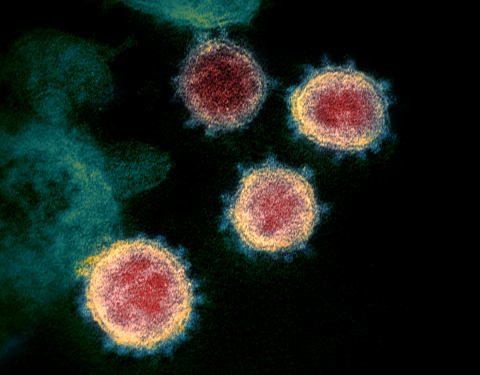
The redox landscape of SARS-CoV-2 infection

Submitted by redoxoma on Wed, 09/16/2020 - 15:09
Sars-Cov-2

Main Article by Flávia Meotti (Instituto de Química da USP) and Francisco R. M. Laurindo (Incor, Faculdade de Medicina da USP)

Post image credits: NIAID / CC BY

The challenge imposed by Covid-19 pandemics has promoted the union of scientists from different fields and technologies, breaking geographic frontiers to solve relevant problems. The focus is, of course, understanding SARS-CoV-2 pathobiology and the ensuing development of improved diagnostic procedures, vaccines and therapeutic compounds. Redox processes certainly belong to this story. In fact, redox pathways can be identified at essentially every level of SARS-CoV-2-related disease and hold a unique potential to integrate knowledge at different systems levels and connect multiple disciplines. Here we briefly highlight, on a somewhat arbitrary and incomplete fashion, redox phenomena of potential significance to this scenario. We hope to inspire our readers to complete this picture and pursue them in depth.

Viral entry into cells: redox and non-redox pathways

The entry of enveloped viruses such as SARS-CoV-2 into cells is a two-step process involving: (1) viral particle binding to cell-surface receptors and (2) fusion of the virion to host cell membranes.

SARS-CoV-2 binding and subsequent entry into the host cell depends essentially on the viral Spike glycoprotein (S glycoprotein). The S glycoprotein forms the characteristic corona of large distinctive spikes in the viral envelope [Xiao et al., 2003]. S glycoprotein contains the S1 domain, a globular region of the protein distal to the virus membrane and the S2 domain, which forms the stalk. S1 domain mediates high-affinity binding with the major (though not exclusive) primary cell receptor, angiotensin-converting enzyme 2, ACE2. Both glycoprotein S and ACE2 exhibit reactive cysteine residues. Membrane fusion is the result of conformational changes in virion envelope depending on its interaction with cells. For SARS-CoV-2, docking to the ACE2 induces conformational changes required for membrane fusion. For enveloped viruses including Newcastle Virus, HIV and many others, thiol/disulfide rearrangements, commented below, are essential for fusogenic activity [Fenouillet et al., 2007; Jain et al., 2008]. For the viral glycoproteins from these viruses, fusion-associated conformational changes are followed and/or preceded by disulfide bond exchanges. This could also be expected for S glycoproteins from both SARS-CoV and SARS-CoV-2, since they contain 39 Cys residues. Following this hypothesis, a series of point mutations replacing cysteines for alanines in S glycoproteins from SARS-CoV revealed that five of seven Cys present at the S1 receptor binding site are essential for association to ACE2 [Wong et al., 2004]. However, the binding of S glycoprotein to ACE2 and infection of mammal cells were largely insensitive to reducing, oxidizing or alkylating agents [Fenouillet et al., 2007; Lavillette et al., 2006]. Rather, for influenza viruses, fusion events have been linked to other stressful events such as cell acidification. These data led to the conclusion that both binding to ACE2 and the fusion capacity of the spike complex are independent of redox switches, in singular contrast to the situation described for a number of other enveloped viruses. However, recent computational simulations indicate that the affinity of SARS-CoV-2 spike protein for ACE2 is strongly impaired when the cysteines present in both proteins (not in only one of them) are in the reduced state, probably due to conformational changes, mainly in ACE2, that affect binding interaction. In contrast, binding is less affected under more oxidizing conditions able to preserve disulfides [Hati & Bhattacharyya, 2020].

Despite being poorly modulated by redox switches, cysteines present at the C-terminal region of the S2 domain undergo post-translational modifications that are crucial to virus infection. Nine of the thirty-nine cysteines in S glycoprotein are clustered between the membrane spanning domain and the C-terminal cytoplasmic tail, with six of these residues being well conserved among different coronaviruses. In SARS-CoV S glycoprotein, the cysteines clustered near to the predicted transmembrane domain were palmitoylated. Mutations of different groups of cysteines significantly decreased protein palmitoylation and virus fusion into mammal cells [Petit et al., 2007]. Since SARS-CoV-2 S glycoprotein shares 79.6% sequence identity with S glycoprotein from SARS-CoV and both contain cysteine clusters at the C-terminal tail, palmitoylation might be important for SARS-CoV-2 fusion as well.

Other viral proteins important for viral replication can exhibit redox modulation. The Nsp9 (non-structural protein 9) from SARS-CoV forms disulfide-linked homodimers important for RNA binding. These dimers assemble in complex quaternary structure. However, cysteine mutations do not impede oligomerization, indicating redundant redox and non-redox mechanisms for RNA binding [Ponnusamy et al., 2008].

Conformational rearrangements of viral envelope glycoproteins by redox reshuffling during virus-cell interaction and fusion have been described in some coronaviruses and many other enveloped viruses, as discussed above. These thiol/disulfide-dependent changes in envelope conformation seem to depend both on autocatalytic processes or cellular thiol oxidoreductases. A relevant redox pathway for the entry of such viruses into cells is the cell surface thiol redox pool of protein disulfide isomerases [reviewed by Tanaka et al., 2020]. The main pool of these abundantly expressed redox chaperones is located at the endoplasmic reticulum, while a small fraction undergoes relocation at the cell surface or is secreted at the extracellular milieu. This so-called pecPDI (peri/epicellular PDI) pool corresponds to ca. 2% of the total pool in endothelial cells [Araujo et al., 2017], however it has been increasingly shown to play important functions in cell adhesion, metalloproteinase regulation, platelet activation, thrombosis and vascular remodeling, among other effects [rev. by Tanaka et al., 2020]. Several evidences indicate that a number of viruses requires pecPDI for cell internalization. Typically, PDI-dependent disulfide reduction of the gp120 viral protein [Fenouillet et al., 2007] or of beta1/beta3 integrins [Wan et al., 2012] mediate viral entry of HIV or dengue viruses, respectively. On the other hand, as commented above, a similar pathway has not been shown for influenza viruses, which do not seem to require pecPDI (nor redox processes at all) for cell internalization. Even so, probably due to additional pathways during viral infection, PDIs have been proposed as therapeutic targets for influenza A and B viruses on the basis of significant prevention of viral replication by a number of compounds known (although nonspecifically) to inhibit PDI, such as juniferdin, 16F16, PACMA31, isoquercetin, epigallocatechin-3-gallate or nitazoxanide. Also, PDI silencing by siRNAs significantly inhibited viral replication [Kim & Chang, 2018]. It is important to note that the concentration of these compounds may be higher than the usual levels that can be attained in vivo and, in addition, SARS Cov-2 was not tested in these studies.

Viral cysteine proteases as potential drug targets

Some other viral proteins with a redox-dependent component have been investigated as putative targets of antiviral drugs. The two cysteine proteases, protease M (Mpro), also referred as 3C-like protease (3CLpro), and the papain-like protease (PLpro) are the main redox dependent anti- SARS-CoV-2 targets studied so far. In these proteases, the catalytic Cys145 (Mpro) or Cys112 (PLpro) exert a nucleophilic attack to the carbonyl group of the scissile peptide bond. The Mpro performs an extensive proteolytic processing of the viral overlapping polyproteins, pp1a and pp1ab, required for viral replication and transcription [Zhou et al., 2020]. The PLpro hydrolyses the non-structural protein (nsp) sequence to the shorter nsp1, nsp2, nsp3 and nsp4 proteins [Han et al., 2005], also essential for viral replication. PLpro can also deubiquitinate or deISGylate host cell proteins, resulting in immune suppression.

Because of its important role in viral cycle and given the absence of closely related homologues in humans, Mpro is an attractive target for antiviral drugs [Pillaiyar et al., 2016]. Alkylation of Mpro Cys145 by Michael acceptors potently inhibits enzyme activity and viral replication in mammal cells [Jin et al., 2020]. Noteworthy, despite the absence of Mpro homologues in humans, covalent bond by Michael acceptors can be quite unspecific and there is a high chance that these compounds affect a broad range of mammal proteins. Therefore, assays that prove target specificity and rule out side effects are imposed. Two relevant limitations for these SARS-CoV-2 assays are the need for a NB3 security level laboratory and the lack of infectivity in wild-type mice, restraining animal models for in vivo studies.

To minimize unspecific and side-effects of thiol-alkylating compounds, some studies have combined virtual screening, structural analyses and functional assays to select compounds that fit and bind with high affinity to the catalytic cleft of these proteases. By using this combination, it was shown that the GC-376, a pre-clinical drug against feline infectious peritonitis [Kim et al., 2016], extensively networks hydrogen bonds with an excellent geometric complementarity to the Mpro active site, making the covalent bond between the GC-376 aldehyde bisulfite and Cys145 [Ma et al., 2020] thermodynamically favorable. The less selective compound disulfiram, clinically used as an anti-alcoholism drug, inhibits both Mpro and PLpro by forming a mixed disulfide between the molecule and the catalytic cysteines [Lin et al., 2018; Xu et al., 2020]. This mechanism can be less efficient since the disulfide bond could be undone by host reducing agents. Disulfiram also forms disulfide bonds with the non-catalytic Cys128 in Mpro and Cys271 in PLpro but with slower kinetics [Lin et al., 2018; Xu et al., 2020]. The compound has an additional mechanism of inhibition by the displacement of the Zn2+ from zinc fingers, altering protein stability [Lin et al., 2018]. α,β-unsaturated esters were also demonstrated to inhibit SARS-CoV-2 PLpro. The nucleophilic attack of the catalytic cysteine forms a covalent thioether with the β carbon [Rut et al., 2020]. Of note, Mpro has eleven other cysteines beyond the catalytic Cys145, but their role in protein structure and function are much less studied, leaving plenty of room for investigations in this field.

Host factor immune response: a plethora of redox pathways

In addition to virus-specific pathways involved in infection, a large number of redox processes — of which we provide only a rough overview —regulate the host immune response at essentially every level.

First, viruses can subvert the host redox environment to their advantage during cell invasion and intracellular replication. A remarkable example is the widely prevalent group of large nucleocytoplasmic DNA viruses (including Poxviruses, Iridoviruses, Mimiviruses and several others). These viruses display Erv-type sulfhydril oxidases, in addition to thioredoxins and in some cases glutaredoxin and other dithiol CysXX(X)Cys enzymes, which together can induce oxidative protein folding in the host cell cytosol [Hakim & Fass, 2010]. In line with the proposed ancestral evolutionary role of these viruses, it is possible that their induced redox protein folding may have predated the oxidative protein folding in the eukaryotic endoplasmic reticulum [rev. by Hakim & Fass, 2010]. Whether similar processes occur for other viruses is unknown.

Some noteful redox-dependent events can regulate immune response to virus infection. Extracellular secretion of low molecular weight thiols such as glutathione (GSH) governs effector T cell responses through decreases in surface redox potentials, the so-called "reductive remodeling strategy" of immune regulation [Yan & Banerjee, 2010]. Similarly, secreted Trx [Plugis et al., 2018] or PDIA1 [Curbo et al., 2009] promote, via disulfide reduction, inactivation or impaired receptor binding of the regulatory cytokine IL-4. In general, cell-surface or extracelllular thiol oxidoreductases can reduce specific thiol targets to activate immune responses [rev. by Tanaka et al., 2020]. In addition, redox-sensitive lysosomal cathepsins are proteases required for SARS-CoV-2 entry. The broad cathepsin inhibitor E64D (inhibits cathepsin B, H, L, and calpain) completely blocked SARS-CoV-2 infection into HEK293 expressing ACE2. A 70% decrease in infection was also achieved by isolated inhibition of cathepsin L, but not of cathepsin B [Ou et al., 2020]. Another interesting protein is GILT (gamma-interferon-inducible lysosomal thiol reductase), which (as its name says) is a dithiol Cys-X-X-Cys-containing protein induced in lysosomes by gamma-interferon during viral infection. GILT was shown to restrict the infection by distinct viruses including SARS-CoV. Interestingly, the restrictive effect was dependent on its lysosomal localization triggered by N-glycosylation, which was abrogated by loss in GILT thiol reductase motifs [Chen et al., 2019]. Moreover, in line with roles of cell-surface protein disulfide isomerases in cell entry, PDIA3 from lung epithelial cells exerts a key role in influenza-A infection by assisting the correct redox folding of viral hemaglutinin during its passage through the host endoplasmic reticulum. Inhibition or silencing of PDIA3 significantly diminishes viral burden and lung immunoinflammatory responses in vivo [Chamberlain et al., 2019].

Last but not least, it is important to point that the above highlights are only a few remarks amid a much more complex redox signaling network spanning the entire immunoinflammatory landscape. This includes many redox-modulated components that will not be covered here such as immune receptors, intracellular post-translational modifications, proteolytic events, transcription factors, gene transcription, extracellular matrix remodeling, cell adhesion, and several others. Likewise, we will not deepen our discussion about Nox NADPH oxidases, only to mention the known important roles of Duoxes in lung epithelial cell defense against influenza A viruses [Vlahos & Selemidis, 2014]. Nox2 has been involved in the formation of neutrophil extracellular traps (NETs), which seem particularly important in the response to SARS-Cov-2 and in the transition from a homeostatic pattern of infection to an uncontrolled inflammatory response [Veras et al., 2020]. Finally, increasing evidence implicate mitochondria in the control of inflammatory cell activation, via redox processes, small intermediates or metabolic reprogramming [Pålsson-McDermott & O’Neill, 2020].

Conclusion and therapeutic perspectives

As briefly discussed here, redox processes involved in viral and more specifically in SARS-CoV-2 infection are complex, multilevel and comprise a number of interconnected oxidizing as well as reducing events. Therefore, it is unlikely that one redox-active compound or intervention will provide a "magic bullet" to mitigate Covid-19. Rather, a mechanism-based target-directed strategy may exploit some "redox Achilles heels" of SARS-CoV-2 at several levels or improve the host inflammatory response. Despite the massive worldwide rush towards drug repurposing, which included a number of redox-active compounds, a good candidate has yet to emerge. Novel redox interventions, therefore, should rely on designing new compounds, peptides, nanobodies or antibodies directed to specific proteins or pathways. Modulation of Nox NADPH oxidases or mitochondrial-associated responses may also contribute to mitigate an inappropriate inflammatory response. Despite these challenges, strategies using small compounds, when used under a rational systems-based approach, might provide interesting perspectives in immune response modulation. A remarkable example is the recent identification in T-cells of >3,000 cysteine residues able to covalently bind natural or newly-designed small electrophiles. Such cysteine targets covered structurally diverse proteins, some with crucial functions in immune response, leading to the identification of several electrophilic compounds able to modulate T-cell activation through distinct mechanisms, highlighting their potential as chemical probes or therapeutic agents [Vinogradova et al., 2020].

As always, successful therapeutic strategies must emanate from basic science studies to reveal in-depth aspects of SARS-CoV-2-related redox pathobiology and cellular/systemic immunoinflammatory responses. Even with the need to urgently fight Covid-19, rigorous preclinical studies on proposed agents cannot be shortcut. The general rule is that the time invested in these studies will pay itself on the long-run in the form of an overall faster track towards secure and effective interventions, even for agents generally regarded as safe.

Francisco R. M. Laurindo¹ and Flávia C. Meotti², Editors
¹Heart Institute (InCor), University of São Paulo Medical School, Brazil
²Institute of Chemistry, University of São Paulo, Brazil


References

  • Araujo, T. L.; Zeidler, J. D.; Oliveira, P. V.; Dias, M. H.; Armelin, H. A.; Laurindo, F. R. Protein disulfide isomerase externalization in endothelial cells follows classical and unconventional routes Free Radical Biology and Medicine, 103: 199–208, 2017.  »  doi: 10.1016/j.freeradbiomed.2016.12.021
  • Chamberlain, N.; Korwin-Mihavics, B. R.; Nakada, E. M.; Bruno, S. R.; Heppner, D. E.; Chapman, D. G.; Hoffman, S. M.; Vliet, A. van der; Suratt, B. T.; Dienz, O.; et al. Lung epithelial protein disulfide isomerase A3 (PDIA3) plays an important role in influenza infection, inflammation, and airway mechanics Redox Biology, 22: 101129, 2019.  »  doi: 10.1016/j.redox.2019.101129
  • Chen, D.; Hou, Z.; Jiang, D.; Zheng, M.; Li, G.; Zhang, Y.; Li, R.; Lin, H.; Chang, J.; Zeng, H.; et al. GILT restricts the cellular entry mediated by the envelope glycoproteins of SARS-CoV, Ebola virus and Lassa fever virus Emerging Microbes & Infections, 8(1): 1511–23, 2019.  »  doi: 10.1080/22221751.2019.1677446
  • Curbo, S.; Gaudin, R.; Carlsten, M.; Malmberg, K.; Troye-Blomberg, M.; Ahlborg, N.; Karlsson, A.; Johansson, M.; Lundberg, M. Regulation of interleukin-4 signaling by extracellular reduction of intramolecular disulfides Biochemical and Biophysical Research Communications, 390(4): 1272–7, 2009.  »  doi: 10.1016/j.bbrc.2009.10.134
  • Fenouillet, E.; Barbouche, R.; Jones, I. M. Cell Entry by Enveloped Viruses: Redox Considerations for HIV and SARS-Coronavirus Antioxidants & Redox Signaling, 9(8): 1009–34, 2007.  »  doi: 10.1089/ars.2007.1639
  • Hakim, M. & Fass, D. Cytosolic Disulfide Bond Formation in Cells Infected with Large Nucleocytoplasmic DNA Viruses Antioxidants & Redox Signaling, 13(8): 1261–71, 2010.  »  doi: 10.1089/ars.2010.3128
  • Han, Y.; Chang, G.; Juo, C.; Lee, H.; Yeh, S.; Hsu, J. T.; Chen, X. Papain-Like Protease 2 (PLP2) from Severe Acute Respiratory Syndrome Coronavirus (SARS-CoV):  Expression, Purification, Characterization, and Inhibition Biochemistry, 44(30): 10349–59, 2005.  »  doi: 10.1021/bi0504761
  • Hati, S. & Bhattacharyya, S. Impact of Thiol–Disulfide Balance on the Binding of Covid-19 Spike Protein with Angiotensin-Converting Enzyme 2 Receptor ACS Omega, 5(26): 16292–8, 2020.  »  doi: 10.1021/acsomega.0c02125
  • Jain, S.; McGinnes, L. W.; Morrison, T. G. Role of Thiol/Disulfide Exchange in Newcastle Disease Virus Entry Journal of Virology, 83(1): 241–9, 2008.  »  doi: 10.1128/jvi.01407-08
  • Jin, Z.; Du, X.; Xu, Y.; Deng, Y.; Liu, M.; Zhao, Y.; Zhang, B.; Li, X.; Zhang, L.; Peng, C.; et al. Structure of Mpro from SARS-CoV-2 and discovery of its inhibitors Nature, 582(7811): 289–93, 2020.  »  doi: 10.1038/s41586-020-2223-y
  • Kim, Y. & Chang, K. Protein disulfide isomerases as potential therapeutic targets for influenza A and B viruses Virus Research, 247: 26–33, 2018.  »  doi: 10.1016/j.virusres.2018.01.010
  • Kim, Y.; Liu, H.; Kankanamalage, A. C. Galasiti; Weerasekara, S.; Hua, D. H.; Groutas, W. C.; Chang, K.; Pedersen, N. C. Reversal of the Progression of Fatal Coronavirus Infection in Cats by a Broad-Spectrum Coronavirus Protease Inhibitor PLOS Pathogens, 12(3): e1005531, 2016.  »  doi: 10.1371/journal.ppat.1005531
  • Lavillette, D.; Barbouche, R.; Yao, Y.; Boson, B.; Cosset, F.; Jones, I. M.; Fenouillet, E. Significant Redox Insensitivity of the Functions of the SARS-CoV Spike Glycoprotein Journal of Biological Chemistry, 281(14): 9200–4, 2006.  »  doi: 10.1074/jbc.m512529200
  • Lin, M.; Moses, D. C.; Hsieh, C.; Cheng, S.; Chen, Y.; Sun, C.; Chou, C. Disulfiram can inhibit MERS and SARS coronavirus papain-like proteases via different modes Antiviral Research, 150: 155–63, 2018.  »  doi: 10.1016/j.antiviral.2017.12.015
  • Ma, C.; Sacco, M. D.; Hurst, B.; Townsend, J. A.; Hu, Y.; Szeto, T.; Zhang, X.; Tarbet, B.; Marty, M. T.; Chen, Y.; et al. Boceprevir, GC-376, and calpain inhibitors II, XII inhibit SARS-CoV-2 viral replication by targeting the viral main protease Cell Research, 30(8): 678–92, 2020.  »  doi: 10.1038/s41422-020-0356-z
  • Ou, X.; Liu, Y.; Lei, X.; Li, P.; Mi, D.; Ren, L.; Guo, L.; Guo, R.; Chen, T.; Hu, J.; et al. Characterization of spike glycoprotein of SARS-CoV-2 on virus entry and its immune cross-reactivity with SARS-CoV Nature Communications, 11: 1620, 2020.  »  doi: 10.1038/s41467-020-15562-9
  • Pålsson-McDermott, E. M. & O’Neill, L. A. J. Targeting immunometabolism as an anti-inflammatory strategy Cell Research, 30(4): 300–14, 2020.  »  doi: 10.1038/s41422-020-0291-z
  • Petit, C. M.; Chouljenko, V. N.; Iyer, A.; Colgrove, R.; Farzan, M.; Knipe, D. M.; Kousoulas, K. Palmitoylation of the cysteine-rich endodomain of the SARS–coronavirus spike glycoprotein is important for spike-mediated cell fusion Virology, 360(2): 264–74, 2007.  »  doi: 10.1016/j.virol.2006.10.034
  • Pillaiyar, T.; Manickam, M.; Namasivayam, V.; Hayashi, Y.; Jung, S. An Overview of Severe Acute Respiratory Syndrome–Coronavirus (SARS-CoV) 3CL Protease Inhibitors: Peptidomimetics and Small Molecule Chemotherapy Journal of Medicinal Chemistry, 59(14): 6595–628, 2016.  »  doi: 10.1021/acs.jmedchem.5b01461
  • Plugis, N. M.; Weng, N.; Zhao, Q.; Palanski, B. A.; Maecker, H. T.; Habtezion, A.; Khosla, C. Interleukin 4 is inactivated via selective disulfide-bond reduction by extracellular thioredoxin Proceedings of the National Academy of Sciences, 115(35): 8781–6, 2018.  »  doi: 10.1073/pnas.1805288115
  • Ponnusamy, R.; Moll, R.; Weimar, T.; Mesters, J. R.; Hilgenfeld, R. Variable Oligomerization Modes in Coronavirus Non-structural Protein 9 Journal of Molecular Biology, 383(5): 1081–96, 2008.  »  doi: 10.1016/j.jmb.2008.07.071
  • Rut, W.; Lv, Z.; Zmudzinski, M.; Patchett, S.; Nayak, D.; Snipas, S. J.; Oualid, F. El; Huang, T. T.; Bekes, M.; Drag, M.; et al. Activity profiling and structures of inhibitor-bound SARS-CoV-2-PLpro protease provides a framework for anti-COVID-19 drug design bioRxiv, 2020.04.29.068890, 2020. Preprint.  »  doi: 10.1101/2020.04.29.068890
  • Tanaka, L. Y.; Oliveira, P. V.; Laurindo, F. R. Peri/Epicellular Thiol Oxidoreductases as Mediators of Extracellular Redox Signaling Antioxidants & Redox Signaling, 33(4): 280–307, 2020.  »  doi: 10.1089/ars.2019.8012
  • Veras, F. P.; Pontelli, M.; Silva, C.; Toller-Kawahisa, J.; Lima, M. de; Nascimento, D.; Schneider, A.; Caetite, D.; Rosales, R.; Colon, D.; et al. SARS-CoV-2 triggered neutrophil extracellular traps (NETs) mediate COVID-19 pathology MedRxiv, 2020.06.08.20125823, 2020. Preprint.  »  doi: 10.1101/2020.06.08.20125823
  • Vinogradova, E. V.; Zhang, X.; Remillard, D.; Lazar, D. C.; Suciu, R. M.; Wang, Y.; Bianco, G.; Yamashita, Y.; Crowley, V. M.; Schafroth, M. A.; et al. An Activity-Guided Map of Electrophile-Cysteine Interactions in Primary Human T Cells Cell, 182(4): 1009–26.e29, 2020.  »  doi: 10.1016/j.cell.2020.07.001
  • Vlahos, R. & Selemidis, S. NADPH Oxidases as Novel Pharmacologic Targets against Influenza A Virus Infection Molecular Pharmacology, 86(6): 747–59, 2014.  »  doi: 10.1124/mol.114.095216
  • Wan, S.; Lin, C.; Lu, Y.; Lei, H.; Anderson, R.; Lin, Y. Endothelial cell surface expression of protein disulfide isomerase activates β1 and β3 integrins and facilitates dengue virus infection Journal of Cellular Biochemistry, 113: 1681-91, 2012.  »  doi: 10.1002/jcb.24037
  • Wong, S. K.; Li, W.; Moore, M. J.; Choe, H.; Farzan, M. A 193-Amino Acid Fragment of the SARS Coronavirus S Protein Efficiently Binds Angiotensin-converting Enzyme 2 Journal of Biological Chemistry, 279(5): 3197–201, 2004.  »  doi: 10.1074/jbc.c300520200
  • Xiao, X.; Chakraborti, S.; Dimitrov, A. S.; Gramatikoff, K.; Dimitrov, D. S. The SARS-CoV S glycoprotein: expression and functional characterization Biochemical and Biophysical Research Communications, 312(4): 1159–64, 2003.  »  doi: 10.1016/j.bbrc.2003.11.054
  • Xu, L.; Tong, J.; Wu, Y.; Zhao, S.; Lin, B. Targeted Oxidation Strategy (TOS) for Potential Inhibition of Coronaviruses by Disulfiram — a 70-Year Old Anti-Alcoholism Drug ChemRxiv,11936292.v1: 2020. Preprint.  »  doi: 10.26434/chemrxiv.11936292.v1
  • Yan, Z. & Banerjee, R. Redox Remodeling as an Immunoregulatory Strategy Biochemistry, 49(6): 1059–66, 2010.  »  doi: 10.1021/bi902022n
  • Zhou, P.; Yang, X.; Wang, X.; Hu, B.; Zhang, L.; Zhang, W.; Si, H.; Zhu, Y.; Li, B.; Huang, C.; et al. A pneumonia outbreak associated with a new coronavirus of probable bat origin Nature, 579(7798): 270–3, 2020.  »  doi: 10.1038/s41586-020-2012-7

Add new comment

A Time for Reinvention

Submitted by redoxoma on Tue, 09/15/2020 - 19:29
Image: Gerd Altmann, by Pixabay

Editor´s page by Francisco R. M. Laurindo and Flávia Meotti

Editorial

Few authors have been – most often indirectly - remembered during the Covid-19 pandemics as much as Gabriel Garcia-Marquez. Now and then we see everyone talking about "times of coronavirus", "times of Covid", "dystopic times", "exceptional times", "difficult times"and so on. As the world was taken by the corona storm, indeed we can talk about different times. Time for protecting ourselves, time for supporting others, time for discovering new challenges about self-isolation, time to turn on a survival mode, time to think collectively. For some of us, time to be healed or –sadly- time to mourn the loss of loved ones. For all, time to be resilient. For us immersed in science, time to fight for research support, time to fight ignorance, time away from the lab, time in the lab with coronavirus-related research, time supporting research agencies, time to talk about science, time for rethinking careers, time to keep the group united at a distance, time for science education...

For sure, each of us has been affected one way or another, most often radically, by this unusual and unexpected sequence of things. The main common denominator in this scenario is that this is a time for reinvention. Just as "doing our best" is to reinvent oneself at every moment, now it is time to rediscover, readapt and reshape ourselves. It is frequently said that the world will come back to the so-called "new normal". It is unclear whether a new normal will indeed be much different from prior times. Rather, the new normal may be more like reinventing peculiar and remarkably individual ways to reach the "old normal". Above all, in this process, this is a time for hope and to employ our science to try not to rely only on hope !

And, as the storm tries to settle, we come back with the Redoxoma Newsletter. And, of course, with a corona article – how could we not think about the related redox aspects? And we also bring (we apologize it is in spanish) the link to an interview with our well-known colleague Prof. Rafael Radi, a redox scientist at the front of Covid-19 fight organization in Uruguay.

Welcome !

The editors

Francisco R. M. Laurindo¹ and Flávia C. Meotti², Editors
¹Heart Institute (InCor), University of São Paulo Medical School, Brazil
²Institute of Chemistry, University of São Paulo, Brazil

Post image from Gerd Altmann by Pixabay licensed as Pixabay License

Add new comment

Editorial

Submitted by redoxoma on Fri, 02/28/2020 - 16:05
Scientist (adapted by Polina Tankilevitch, from Pexels)

Editor´s page by Francisco R. M. Laurindo and Flávia Meotti

New faces are a feature of our new Redoxoma Newsletter. We start with the emerging biochemistry of persulfides and hydrogen sulfide and follow on the entirely novel concept of peroxymonocarbonate as a mechanism of peroxiredoxin hyperoxidation in the presence of bicarbonate/ carbon dioxide. And we go on in Shakespeare´s style from "something rotten in the redox kingdom" to "to adhere or not to adhere: this is the question". I believe Prince Hamlet would have loved the dualities of redox biochemistry - as confronted to the dualities of life. Speaking of dualities, we come back once more to scientific publications, with an important statement summary from our colleagues. And we end with a translational dive into the redox pathways of sickle cell anemia. These brief notes highlight the multidisciplinary mood of Redoxoma investigators and the enthusiastic pace of research being developed in the network.

Welcome to the novel issue of the Redoxoma Newsletter !

Francisco R. M. Laurindo¹ and Flávia Meotti², Editors
¹Heart Institute (InCor), University of São Paulo Medical School, Brazil
²Institute of Chemistry, University of São Paulo, Brazil

Add new comment

The evolution of Redoxoma Word Cloud: an essay on [not-that-] useless trivia

Submitted by redoxoma on Mon, 09/30/2019 - 16:23
CEPID Redoxoma wordcloud

Redoxoma Highlights, by Francisco R. M. Laurindo
Corresponding author e-mail: francisco.laurind@hotmail.como@hc.fm.usp.br

As scientists, likely we would love to be what we think, but for practical purposes we are what we write. Wouldn't that be interesting to have a big picture of what words we have been writing in Redoxoma? Obviously, computing this is no easy task, but at least looking at the titles of our publications could give us a hint of which are our favorite subjects. Reporting word statistics would certainly be boring, however word clouds can provide quantitative and unbiased yet understable estimates. After all, if it is true that a picture is worth a thousand words, one should be able to learn something from a picture made of words.

With this idea in mind, I took the titles of all papers published in our Cepid-Redoxoma (from our site) an grouped them in 3 datasets: 2013 to 2015, 2016 to 2017 and 2018 to mid-2019. The aim was to contribute to those collections of amusing and [not-that-] useless trivia.

Obviously the datasets are not quantitatively equal, but doing some experimentation with distinct modes of organization did not yield relevant alterations in the overall results. Word Clouds were constructed using WordItOut software, organizing the word size by frequency. Results are shown in the figures below. As expected, a few bits of information can be derived from them. Here I bring a few interesting highlights and invite our readers to add additional observations in our site !

  • Our main keywords are quite conserved across the years. This indicates a consistency of the central interests and research tracks, which I see as a faithful portrait of Redoxoma: focus on solid questions away from fashionisms. The risk of a lack of innovative and refreshing ideas, I believe, is readily dismissed by a closer exam of the published articles.
  • Clearly, "mitochondrial" is the outstanding keyword in all datasets (we all know who is responsible for that...) no matter how we group the data. Recently, however, this remarkable organelle has been challenged by "oxygen"- quite a fair game I would say, given the true respectability of the basic element our work is based on.
  • The words indicate we are definitely a protein rather than a gene group. Among the proteins, thiol proteins are steadily prominent: peroxiredoxins (Prx), isomerase (which derives from the PDI works, no personal bias please), Ohr, etc.
  • Oxidant-related wording (oxidant, oxidative, disulfide, peroxynitrite) quite clearly predominates over reductant-related wording. This suggests that oxidation still dominates the collective uncounscious of redox investigators. Maybe it would be a good time to focus on reducing pathways?
  • Some words are notable for their absence or paucity: the top candidate to me is "antioxidant".
  • It was reassuring that manicheist jargon, such as "deleterious", "protective", "defense", "damaging", "toxic", etc does not dominate our wording. This reflects a state-of-the-art approach to free radical-dependent biology and medicine, in which thoughts are driven towards mechanisms rather than good-or-bad classifications.
  • The words tell us we definitely seem to focus more on death rather than survival. And stress of course. Not surprising.
  • I was surprised that "singlet" consistently outperformed "peroxide", "superoxide" and related words. Similarly, "photodynamic", not a usual word in redox biomedicine, was consistently high accross all datasets. Congratulations to all those involved.
  • Copper was the most popular metal in the two earliest datasets, but now it has been surpassed by iron, with zinc and selenium as emerging runnerups. An interesting competition to watch.
  • Not unexpectedly, there is good competition between methods/chemistry-related keywords and application/physiology ones. Will [spectroscopy, electrochemical, mass, quantification, activities, synthesis, reactions, adduct, etc...] outcompete [metabolic, vascular, aortic, heart, neurodegeneration, olfactory, caloric, etc...] ? The jury is out, but my personal veredict is that we should switch the word "competition" to "balance".
  • "Human" stands nearly at the same rank as "rats" , but both are largely surpassed by "cell(s)". Also not surprising for a mechanistic group.
  • The most recent dataset shows a wellcome emergence/increase of some interesting terms: "structural", "molecular", "model" and "mechanism". Hopefully we will keep writing more about these.
  • In a context for the ugliest words, my vote would stay with "characterization" and "effects", which are description-related terms.
  • The words tell us that for sure we kept doing many "novel" and "new" things at Redoxoma.

It is important to say that these exercises have several intrinsic biases from the scientific standpoint and should not be taken as a picture of the science we do at Redoxoma. Hopefully, however, these diagrams will give us some material for fueling our conversations and maybe some thoughts. And some fun, of course.


Francisco R. M. Laurindo, Editor in Chief of Redoxoma Newsletter at Heart Institute (InCor),
University of São Paulo Medical School, Brazil


Add new comment

Editorial

Submitted by redoxoma on Mon, 09/30/2019 - 15:47
CEPID Redoxoma

Editor's page by Francisco R. M. Laurindo and Flávia Meotti

Time keeps going fast and 2019 advances quickly to its last third. So do our investigations, as you can see in this new issue of the Redoxoma Newsletter!

We start on the bright side of things, with a nice perspective on fluorescent probes to assess redox processes in biological systems, written by Dr. Marcelo Comini, our colleague from Uruguay.

We follow on form and function: a great highlight on the interplay between mitochondrial dynamic morphology and calcium uptake.

Then you should more than just say NO and read the highlight depicting innovative pathways of nitric oxide metabolism involving dinitrosyl iron complexes.

And we finish with a puzzle on useless trivia: which is the most frequent word in the title of Redoxoma papers? Read this essay to discover.

Francisco R. M. Laurindo¹ and Flávia Meotti², Editors
¹Heart Institute (InCor), University of São Paulo Medical School, Brazil ²Institute of Chemistry, University of São Paulo, Brazil

Add new comment

Editorial

Submitted by redoxoma on Wed, 05/15/2019 - 17:42
Foto: Marcos Santos/USP Imagens (sob licença CC BY-NC 3.0 BR)

Editor's page by Francisco R. M. Laurindo and Flávia Meotti

Welcome to our new issue of the Redoxoma Newsletter!

Challenges. We start with a frank talk by a young colleague which we believe will be quite appealing to our readers, both the youngs who face career challenges and the experienced investigators who face the challenge of supervising post-docs who face career challenges. Quite an important issue in our view and a major challenge for Brazilian science.

And we keep on challenges. This time a radical connection on the pathways of peroxynitrite reactivity and decomposition, involving the carbonate radical, very much an original Redoxoma contribution !

And we keep on original Redoxoma contributions. This time off the beaten track, on singlet oxygen and its implication on sepsis and inflammation.

And we keep on inflammation. This time discussing how neutrophils keep their balance and sum up on paradigms of redox signaling.

And we finish with a “D”. We invite to discover how.

Francisco R. M. Laurindo¹ and Flávia Meotti², Editors
¹Heart Institute (InCor), University of São Paulo Medical School, Brazil ²Institute of Chemistry, University of São Paulo, Brazil

Post image credits: Marcos Santos/USP Imagens under CC BY-NC 3.0 BR license.

Add new comment

Editorial

Submitted by redoxoma on Thu, 02/28/2019 - 18:16
Welcome to the CEPID Redoxoma Newsletter

Editor's page by Francisco R. M. Laurindo and Flávia Meotti

Welcome to the new issue of our Redoxoma Newsletter!

Soon you will notice a nice feature of this issue: an essay on electronic laboratory notebooks, which are coming steadily to replace the paper notebooks. Or not? Take your own conclusions, there many issues worth discussing. But the best part, in my view, is that the essay was conceived and written by two PhD students from Redoxoma. Hopefully this will be a good example to be followed by other PhD students and post-docs: we are eagerly waiting for your contribution!

And we follow with nice works on how reactive intermediates from Nox1 NADPH oxidas/e are regulated by protein disulfide isomerase. And the story goes on with a critical appraisal of the role of the proteasome main particle on physiological protein degradation. Works that explore the mechanistic vibe of Redoxoma. And from mechanisms to application as our main characteristic, we present the Innovation Corner with some advances in the I of RIDC (or Cepid).

Finally, more good news. We have significantly expanded the Newsletter diffusion range and hope the newcomers will enjoy staying with us.

Francisco R. M. Laurindo¹ and Flávia Meotti², Editors
¹Heart Institute (InCor), University of São Paulo Medical School, Brazil
²Institute of Chemistry, University of São Paulo, Brazil

Add new comment

Editorial

Submitted by redoxoma on Fri, 12/07/2018 - 13:37
Ed. December, 2018

Editor's page by Francisco R. M. Laurindo and Flávia Meotti

We have a varied menu to fulfill our scientific curiosity starting with a discussion on lipid redox code and the amazing variety of these compounds, which is just beginning to be scratched from the surface, as further exemplified by a Redoxoma work describing protein aggregation induced by lipid oxidation-derived aldehydes. We follow on interesting discoveries related to uric acid hydroperoxide functions.

The rest goes from fun to serious discussions, just as Science itself. We bring a list of the most popular redox genes, which you will find amusing and thought-provoking. The serious part follows our previous issues involving scientific publishing practices to discuss inconvenient but necessary thoughts about publications. Just as a reminder, the oppinions expressed in our articles represent the author's views and not those from Redoxoma.

Welcome to our Redoxoma Newsletter issue.

Francisco R. M. Laurindo¹ and Flávia Meotti², Editors
¹Heart Institute (InCor), University of São Paulo Medical School, Brazil
²Institute of Chemistry, University of São Paulo, Brazil

Add new comment

More on impact factor metrics: are we ready to get rid of them?

Submitted by redoxoma on Wed, 09/26/2018 - 21:20
Paper sheets

The Radical-Free corner by Francisco R. M. Laurindo

Recently, a young investigator wrote to Nature [1] urging to "stop saying that publication metrics do not matter and tell early-career researchers what does" when rating the scientific achievements of young investigators. This message highlights that increasing awareness against the inappropriate use of impact factor (IF) metrics for evaluating CVs is bringing, as a side effect, undertainty and lack of clarity on how someone's career achievements will be evaluated. This boils down to the simple question of whether we are ready to get rid of IF metrics.

First of all, it is important to say that it is increasingly accepted that using IFs as the sole metrics to evaluate someones's achievements in science is flawed by a number of reasons [2]. This tendency has led to the San Francisco "Declaration on Research Assessment" (DORA) in 2012, which has been since signed by over 500 institutions and 12000 individuals, calling among other issues, to "the need to eliminate the use of journal-based metrics, such as Journal IFs, in funding, appointment, and promotion considerations" and " the need to assess research on its own merits rather than on the basis of the journal in which the research is published ". In fact, recent experiences indicate that hiring investigators on the basis of addressing their achievements and contributions with interviews rather than traditional publication-based CVs has led to improved results [3]. Indeed, the current NIH-type biosketch (not dissimilar to FAPESP) is centered on the value of each individual's contribution to science.

All these welcome advances may have raised the perception, expressed in the comment from the first paragraph, that IF-related metrics do not matter any more. I believe the cold fact is that they still do to a reasonable extent and presently there is no completely adequate replacement for them, including the evaluation of young investigators. The distortions of strictly adhering to IFs and numeric scores should not be taken to mean that we have clear validated alternative methods available. Looking to someone's specific contributions and having a holistic approach when comparing a few candidates for academic purposes may indeed prove successful even with today's tools due to the low scale of this task. But even so, many evaluators still run their parallel evaluations of IF metrics, as this is yet so much embedded into our collective unconscious and provides some numerical scores with a security blanket of objectivity. However, problems become particularly acute when competitively evaluating a large number of CVs, e.g., regarding scholarships or large-scale research awards. As unfair and innaccurate as it would be to blindly rely only on the metrics, it would also be unfair to ignore them. Despite the limitations, there is indeed some gross correlation at least between the highest journal IFs with the quality and completeness of published work and ensuing amount and quality of the effort put into it. Along this line, IFs and number of articles do tell something about the capacity of the individual to choose important problems, to focus deep into a given problem until the end, to work hard and intensively into scientific questions and to be able to finish coherent stories about them. For the early-career investigator, relying solely on citations can be innappropriate because many good articles take a long time to get cited. Thus, number of published articles and their IF-related metrics are still a default basis for early-career CV evaluation. In fact, much before IFs were invented, everyone knew the most prestigious publications and those who published on them were positively considered.

On other hand, the limitations of those metrics are real. I believe that down the road these numbers can only help separate candidates that are very good or excellent from those that are merely good or median. However, metrics can significantly fail when trying to separate the top candidates from those that are just not as excellent. So, what can be done to improve on these issues? The DORA followers are getting away with all the metrics, however this leaves everyone, specially the young investigators, uneasy about how they will be evaluated and what are the best career strategies [1]. Without having the illusion (or arrogance?) that I will set the last word on this complex subject, I believe we are not yet ready to get rid of IF metrics in general, but the system can indeed take several extra paths to perfect, reinterpret and at the end eventually ignore them. Here I list some features that are increasingly being taken into account in the evaluation of young investigator's CVs. In the absence of a better collective term, I will call them modifiers, that is, each of them can potentially enhance or decrease potential inferences derived from metrics.

An important modifier is the intrinsic quality of the work, that is, overall degree of innovation, extent of contribution, implications for novel ideas or for potential applications, accuracy and completeness of the investigative strategy, and so on. Logically, the works having these qualities will usually take much more time to be performed and this allows less time to publish other papers, potentially decreasing the number of published items. Additionally, in some cases, these works may be published in journals that despite their solid reputation and tradition, along with lengthy and demanding reviews, do not display proportionally high IFs. Such are the cases of Journal of Biological Chemistry, Journal of Molecular Biology, American Journal of Physiology… among others. Contrarily, some journals use a number of strategies to unrealistically maximize their IFs and the intrinsic value of the work they publish may not be proportionally as high (this is a good theme for future discussions). Evaluators should take all these issues into account. However, it is important that the scientist being evaluated does not assume that reviewers will appreciate the quality of someone's work by default: reviewers are uniformely very busy and may not be from the particular subarea that would readily understand the specifics. Thus, the intrinsinc qualities of each one's work have to be explicitly clarified by the author. The investigator's biosketches from many research agencies, including Fapesp, provide appropriate space for the investigator to write in a few lines what is the contribution and novelty of that paper, as well as anything else that can indicate intrinsic value (e.g., it is a pioneer work in specified aspects, it contributed to diagnostic or therapeutic advances, it served as a basis for public policies, etc) or the community's perception of value (e.g., it promoted the invitation for a relevant talk, it was the theme of an editorial comment or chosen as the cover article, etc). This will contribute to identify intrinsic qualities in published work, which will adjust and improve the interpretations of numeric scores, in some cases even allowing one to get totally out of them. Interestingly, for unclear reasons, investigators have rarely made use of this strategy at Fapesp, although the biosketch format allows that.

On the opposite side, in some cases young investigators display a CV characterized by a large number of publications, however of intrinsic low value: incremental contributions, not-so innovative advances or questionable methodology. Given the conundrums of the scientific publishing scenario nowadays, these works do get published somehow. In other cases, the works are multiauthored without a clear contribution of the author being evaluated, which sometimes will appear as a middle-author amongst several others. There is nothing wrong – and actually it is good – to get involved in many investigations from a given group. Moreover, in some cases of multiple high-quality cooperative work, a middle position by no means indicates a negligible contribution. Again, disclosing the author's contribution for that particular work in the space provided in the biosketch is essential and will help understand the potential value of the author's contribution and how it differs from the so-called "salami-splicing" type of CV. Moreover, I suggest that the authors separately highlight, in their biosketches, only their few principal works by which they want to be evaluated and leave the others as a group of collaborations. That will avoid that noise from too many works obscures what really matters.

A further enhancer in a CV is what I would call "vertical coherence", that is, the connectivity accross each of the investigator's papers, allowing one to foresee the emergence of an investigative track. This multiplies the importance of each work, so their overall value is larger than the sum of each part. Again, these connections must be emphasized and explained by the investigator.

Another relevant aspect is that there are other dimensions of impact of a scientific work that transcend the scientific sphere. This is recognized now by many research agencies, including Fapesp, and comprise : 1) Social relevance and 2) Economic impact. Furthermore, in some areas, general metrics of impact are lower than those in other areas as a characteristic of the field. Again, in such cases, the relevant information will not be readily obvious for reviewers that are not too specialized and thus should be clearly highlighted by the investigator.

These considerations indicate that a number of parallel aspects can affect the perception and interpretation of the IF metrics, providing a more accurate and fair picture. Are these modifiers subjective? Perhaps yes to a good extent, but one has to balance the problems. Certainly, at this time we still face a paradox. As discussed above, IF metrics is still embedded into the system. On the other hand, it is likely possible that incorporating the modifiers described in this essay and trying more and more to have a systematic approach to them will enhance the capacity to select the best achievements while getting off the numerical score tiranny. Decorating the basic metrics with such a "systematic subjectivity" evaluation and perfecting it along time seems more realistic, feasible and less traumatic to early-career scientists than just abandoning metrics all of a sudden. While we love to hate IFs, they are still deep into our minds.


  1. J. Tregoning. How will you judge me if not by impact factor? Nature, 558(7710): 345, 2018 | doi: 10.1038/d41586-018-05467-5
  2. J. K. Vanclay. Impact factor: outdated artefact or stepping-stone to journal certification? Scientometrics, 92(2): 211-38, 2011 | doi: 10.1007/s11192-011-0561-0
  3. S. L. Schmid. DORA Molecular Biology of the Cell, 28(22): 2941-4, 2017 | doi: 10.1091/mbc.e17-08-0534

Francisco R. M. Laurindo, Editor in Chief of Redoxoma Newsletter
Heart Institute (InCor), University of São Paulo Medical School, Brazil


Add new comment