Redoxoma Highlights

Revealing the cross-talk between nitric oxide metabolites

Submitted by redoxoma on Mon, 09/30/2019 - 15:44
Artistic EPR spectra

Redoxoma Highlights, by Daniela Truzzi
Corresponding author e-mail: dtruz@hotmail.comzi@iq.usp.br

Nitric oxide (NO) is an endogenously produced diatomic radical that regulates fundamental biological functions. Although NO is a free radical, its reactivity in biological media is selective toward other radicals and transition metal centers. NO metabolites include S-nitroso thiols (RSNOs), nitrite, peroxynitrite, nitrosylated heme proteins, and dinitrosyl-iron complexes (DNICs). Among these metabolites, RSNOs have gained considerable attention due to their possible involvement in NO signaling. Biological formation of RSNO can occur by reaction of thiols with N2O3, peroxynitrite, other S-nitroso thiols (transnitrosation reactions), nitrosylated heme proteins and by a direct reaction between thiyl radicals (RS) and NO. Since most of these reactions are either slow or have low specificity for a signaling process, it has been proposed that S-nitrosation involves transfer of NO from DNICs to biothiols. DNICs are important NO-metabolites, which are able to trigger vasodilation, to inhibit platelet aggregation and skin wound healing. Nevertheless, little is known about the dynamics of DNICs generation under physiological conditions. By analyzing DNIC assemble from the reaction between NO, Fe(II) and low molecular weight biothiols (cysteine and glutathione) in aqueous media, pH 7.4, we detected mono-nitrosyl iron complex intermediate(s) and thiyl radicals (RS) as co-products. By demonstrating that formation of DNICs yields RS in a NO rich environment, these results provide a novel route for S-nitroso thiol formation in biological media. Additionally, this study explains previous reports showing that DNICs and RSNOs are simultaneously formed in macrophages exposed to NO. If such mechanism favors certain biothiols in forming RS and thus provides specificity to RSNO formation, remains an open question. Further studies of DNICs assembly with different biothiols may contribute to answer it.

Importantly, different biothiols may react through distinct mechanism with DNICs, opening the question if all biothiols would similarly have RS as an intermediate. This is of high relevance when considering signaling pathways and specificity.

This study involved collaboration between researchers from CEPID Redoxoma and from University of California, Santa Barbara.


Related article:

  1. D. R. Truzzi, O. Augusto, P. C. Ford. Thiyl radicals are co-products of dinitrosyl iron complex (DNIC) formation Chemical Communications, 55(62): 9156–9, 2019. | doi: 10.1039/c9cc04454j

Daniela Truzzi, from Department of Biochemistry,
Institute of Chemistry, University of São Paulo, Brazil
Add new comment

A link between mitochondrial shape and function in Ca²⁺ signaling

Submitted by redoxoma on Mon, 09/30/2019 - 13:24
Calcium

Redoxoma Highlights by Sergio Menezes and Alicia Kowaltowski
Corresponding author e-mail: alicia@kowaltowski@iq.usp.br

Differently from what one might expect from the textbook representations, mitochondria are not static organelles which are always in well-defined round shapes. In fact, they are highly dynamic organelles which undergo constant cycles of fission and fusion with nearby mitochondria [1]. “mitochondria are not static organelles … in well-defined round shapes” The balance between these fusion and fission events within the cell is what will define the shape of the mitochondrial network, which can range all the way from several separate round shaped mitochondria (high fission, low fusion balance) to very interconnected networks of elongated mitochondria that span throughout the cell (high fusion, low fission balance) [1]. Several factors like cell type, nutrient availability, progression through the cell cycle and even pathological conditions contribute to this fusion/fission balance within the cell [2, 3]. The study of these dynamic modulations of mitochondrial morphology is broadly called "mitochondrial dynamics" [1], and is a field that has received a lot of attention in the last two decades.

In our recent study accepted for publication in The Faseb Journal [preprint available at https://www.biorxiv.org/content/10.1101/624981v1], we show a novel role for mitochondrial dynamics in regulating cellular Ca2+ signaling and homeostasis. “we show a novel role for mitochondrial dynamics in regulating cellular Ca2+ signaling and homeostasis” Mitochondria forced into a pro-fusion phenotype through the inhibition of the fission protein DRP1 (by competition with a dominant-isoform) presented increased mitochondrial Ca2+ uptake rates and maximal uptake capacity, while mitochondria forced to a fission phenotype by the knockdown of the fusion protein MFN2 showed the opposite effect. Mitochondrial Ca2+ uptake is a process mediated by the entry of Ca2+ ions in the mitochondrial matrix through the protein MCU (mitochondrial calcium uniporter) driven by the negative-inside mitochondrial membrane potential, and has been extensively shown to impact several processes involving Ca2+ signaling in the cell [6]. In our work we also show that one of these regulated Ca2+ uptake processes, known as store-operated Ca2+ entry (a homeostatic mechanism by which cells are capable of replenishing their ER Ca2+ stores by promoting extracellular Ca2+ entry through the membrane) is modulated by mitochondrial morphology, with more fragmented mitochondria resulting in an impairment of this process, while more fused mitochondria resulted in a faster activation of extracellular Ca2+ entry. We also have observed a reduction in basal cytoplasmic and ER Ca2+ levels in the cells with more fragmented mitochondria, which was associated with increased levels of ER stress markers, showing that mitochondrial morphology can also regulate these aspects of cellular Ca2+ homeostasis.

By showing that the modulation of mitochondrial morphology can impact mitochondrial Ca2+ uptake and promote changes in Ca2+ homeostasis in the cell, this work establishes a new connection between mitochondrial dynamics and cell signaling.


References

  1. L. Tilokani, S. Nagashima, V. Paupe, J. Prudent. Mitochondrial dynamics: overview of molecular mechanisms Essays In Biochemistry, 62(3): 341–60, 2018. | doi: 10.1042/ebc20170104
  2. M. Liesa, O. Shirihai. Mitochondrial Dynamics in the Regulation of Nutrient Utilization and Energy Expenditure Cell Metabolism, 17(4): 491–506, 2013. | doi: 10.1016/j.cmet.2013.03.002
  3. R. Horbay, R. Bilyy. Mitochondrial dynamics during cell cycling Apoptosis, 21(12): 1327–35, 2016. | doi: 10.1007/s10495-016-1295-5
  4. M. F. Forni, J. Peloggia, K. Trudeau, O. Shirihai, A. J. Kowaltowski. Murine Mesenchymal Stem Cell Commitment to Differentiation Is Regulated by Mitochondrial Dynamics Stem Cells, 34(3): 743–55, 2015. | doi: 10.1002/stem.2248
  5. M. Vig, J. Kinet. Calcium signaling in immune cells Nature Immunology, 10(1): 21–7, 2008. | doi: 10.1038/ni.f.220
  6. A. Spät, G. Szanda, G. Csordas, G. Hajnóczky. High- and low-calcium-dependent mechanisms of mitochondrial calcium signalling Cell Calcium, 44(1): 51–63, 2008. | doi: 10.1016/j.ceca.2007.11.015

Sergio Menezes and Alicia Kowaltowski, from Department of Biochemistry,
Institute of Chemistry, University of São Paulo, Brazil

Add new comment

The interaction between peroxides and the labile iron pool: a new side of the coin

Submitted by redoxoma on Thu, 09/27/2018 - 15:51
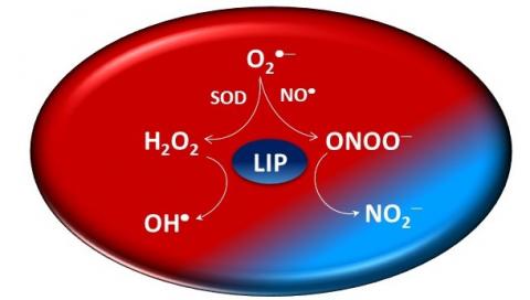
LIP, by J. Toledo Jr. et. al
The redox iron-fist of cells may not always be so harsh, this study suggests.

Redoxoma Highlights by José Carlos Toledo Jr.

Every cell keeps low levels of an iron source called the Labile Iron Pool (LIP), which serves primarily as a reservoir of iron to be loaded into apo iron-proteins. LIP iron is probably weakly bound to proteins and small molecules, but its precise nature and cellular location are not yet known. LIP quantity varies among different cell types and is tightly regulated by complex homeostatic mechanisms, deregulation of which can be dangerous. For instance, aberrant accumulation of LIP is a hallmark of cardiovascular diseases and neurological disorders. Although still not clear, it is widely accepted that, upon a reaction with the ubiquitous hydrogen peroxide, LIP facilitates the production of highly reactive species that oxidize proteins, DNA and membrane lipids, and that these damages contribute to multiple pathologic conditions. In agreement, in vitro, iron reduces hydrogen peroxide to the hydroxyl radical, a very reactive oxidant species. From this perspective, it makes sense that normal cells keep low levels of LIP, which presumably minimizes production of oxidants. However, our recent finding suggests there is another side of this story. It was recently found that LIP reacts with a peroxide called peroxynitrite, a toxic oxidant that is formed in cells, and thereby actually decreases peroxynitrite-dependent oxidation of an intracellular indicator. This observation may change the widespread notion that the LIP is solely a cellular pro-oxidant iron source and provides additional information to understand biological phenomena in conditions where peroxynitrite and LIP overlap.


Related article:

  • F. C. Damasceno, A. L. Condeles, A. K. B. Lopes, R. R. Facci, E. Linares, D. R. Truzzi, O. Augusto, J. C. Toledo. The labile iron pool attenuates peroxynitrite-dependent damage and can no longer be considered solely a pro-oxidative cellular iron source Journal of Biological Chemistry, 293(22): 8530–42, 2018 | doi: 10.1074/jbc.ra117.000883

José Carlos Toledo Jr., PhD. Professor at Department of Chemistry,
Faculdade de Filosofia, Ciências e Letras de Ribeirão Preto, University of São Paulo, Brazil


Add new comment

Ohr (Organic Hydroperoxide Resistance Proteins) is central in the response of bacteria to oxidants released during inflammatory processes

Submitted by redoxoma on Thu, 09/27/2018 - 15:37
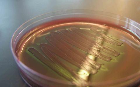
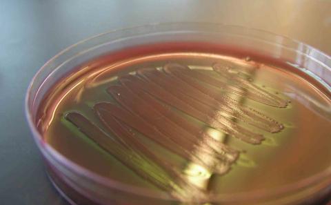
A Petri Plate with E. coli growing (by www.biology101.org)
This cysteine-based peroxidase unique to prokaryotes may provide relevant clues for designing novel anti-bacterial therapies.

Redoxoma Highlights by Luis. E. S. Netto

Oxidants play central roles in cell signaling during host-pathogen interactions. Among them, hydroperoxides of arachidonic acid are mediators of inflammatory processes in mammals, whereas hydroperoxides of linoleic acid play equivalent roles in plants. Therefore, hydroperoxide levels are strictly controlled by both hosts and pathogens. CEPID redoxoma is leading the biochemical/structural characterization of bacterial Ohr (Organic Hydroperoxide Resistance Protein), which is an unique Cys-based peroxidase with very distinct features than analogous proteins from host organisms [1, 2, 3].

Recently, we showed that Ohr reduces fatty acid hydroperoxides and peroxynitrite at extremely high rates [4]. Furthermore, bacterial mutants devoid of Ohr gene display high sensitivities to these oxidants, whereas mutant strains deficient for other peroxidases were equally sensitive than wild type cells to fatty acid hydroperoxides. Therefore, Ohr plays central roles in the bacterial response to two hydroperoxides that are at the host–pathogen interface [4]. Ohr enzymes were previously thought to be restricted to prokaryotes, but we found Ohr orthologues also in eukaryotic microorganisms (some pathogenic), many of them located in mitochondria [5].

Our group also described novel findings related to OhrR, which is a transcriptional regulator that represses Ohr expression. We recently showed OhrR also represses diguanylate cyclase expression in Chromobacterium violaceum. Furthermore, deletion of OhrR attenuated the virulence of C. violaceum in mice, decreasing the bacterial burden in the liver [6]. Since Ohr-OhrR system is absent in both mammals and plants, it represents an attractive target for drug development.


References

  1. J. R. R. Cussiol, T. G. P. Alegria, L. I. Szweda, L. E. S. Netto. Ohr (Organic Hydroperoxide Resistance Protein) Possesses a Previously Undescribed Activity, Lipoyl-dependent Peroxidase Journal of Biological Chemistry, 285(29): 21943–21950, 2010 | doi: 10.1074/jbc.m110.117283
  2. M. A. Oliveira, B. G. Guimarães, J. R. Cussiol, F. J. Medrano, F. C. Gozzo, L. E. Netto. Structural Insights into Enzyme–Substrate Interaction and Characterization of Enzymatic Intermediates of Organic Hydroperoxide Resistance Protein from Xylella fastidiosa Journal of Molecular Biology, 359(2): 433–445, 2006 | doi: 10.1016/j.jmb.2006.03.054
  3. E. Piccirillo, T. G. P. Alegria, K. F. Discola, J. R. R. Cussiol, R. M. Domingos, M. A. de Oliveira, L. de Rezende, L. E. S. Netto, A. T. Amaral. Structural insights on the efficient catalysis of hydroperoxide reduction by Ohr: Crystallographic and molecular dynamics approaches PLOS ONE, 13(5): e0196918, 2018 | doi: 10.1371/journal.pone.0196918
  4. T. G. P. Alegria, D. A. Meireles, J. R. R. Cussiol, M. Hugo, M. Trujillo, M. A. de Oliveira, S. Miyamoto, R. F. Queiroz, N. F. Valadares, R. C. Garratt, et al. et al.. Ohr plays a central role in bacterial responses against fatty acid hydroperoxides and peroxynitrite Proceedings of the National Academy of Sciences, 114(2): E132–E141, 2016 | doi: 10.1073/pnas.1619659114
  5. D. Meireles, R. Domingos, J. Gaiarsa, E. Ragnoni, R. Bannitz-Fernandes, J. da Silva Neto, R. de Souza, L. Netto. Functional and evolutionary characterization of Ohr proteins in eukaryotes reveals many active homologs among pathogenic fungi Redox Biology, 12: 600–609, 2017 | doi: 10.1016/j.redox.2017.03.026
  6. M. Previato-Mello, D. de A. Meireles, L. E. S. Netto, J. F. da Silva Neto. Global Transcriptional Response to Organic Hydroperoxide and the Role of OhrR in the Control of Virulence Traits in Chromobacterium violaceum Infection and Immunity, 85(8): , 2017 | doi: 10.1128/iai.00017-17

Luis. E. S. Netto, PhD. Professor at Department of Genetics and Evolutionary Biology,
Institute of Biosciences, University of São Paulo, Brazil


Add new comment

The mechanism of photo-induced membrane leakage

Submitted by redoxoma on Thu, 09/27/2018 - 11:42
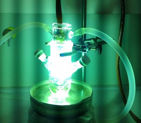
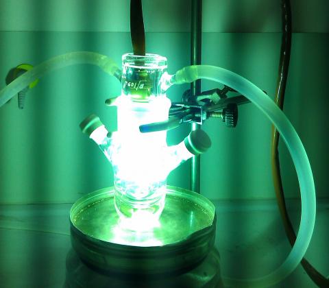
Philips HPK125 UV lamp in a photochemical immersion well reactor 50 mL
Membrane permeabilization by light involves contact between photosensitizer and the lipid double bond, yielding membrane-disrupting aldehydes

Redoxoma Highlights by Mauricio da S. Baptista

Light is fundamental for life, making feasible processes such as photosynthesis, and vision. Photodynamic therapy is a clinical modality able to treat a variety of diseases with light. However, light it is also responsible for spoiling foods and for the development of skin cancer, due to photosensitization reactions, in which molecules transform light energy into chemical reactivity, very frequently causing oxidations. Our group has accumulated evidence that membranes are key targets of the photo-induced reactions. The general mechanisms of lipid photo-oxidation have been known for a long time, but oxidation by singlet oxygen, which is a diffusing molecule, does not suffice to explain the known fact that photosensitizers that bind to membranes are more effective in making them permeable and therefore in destroying cells. We designed experiments to measure efficiency of membrane leakage by photosensitizers delivering almost the same amount of singlet oxygen to the membranes, but having a significant difference in the extent of the direct physical contact with lipid double bonds. We also identified and quantified all products generated by photosensitizers, such as hydroperoxides, alcohols, ketones and phospholipid aldehydes. We found that there always is significant accumulation of truncated lipid aldehydes in leaking membranes. Another important result was that permeabilization of membranes was invariably coupled to photobleaching, i.e., the photosensitizer gets degraded. Therefore, if one wants to have a more effective photosensitizer, we have to find new ways to regenerate it. Also, as a strategy for developing more efficient sunscreens, we need to create a way to prevent aldehydes from building up. This study was carried out as a doctoral project of Isabel Bacellar, the first author of the article, and involved the collaboration of CEPID researchers Redoxoma Paolo Di Mascio and Sayuri Miyamoto. Ronei Miotto and Rodrigo Maghdissian Cordeiro (Federal University of ABC), Professor Gonzalo Cosa (McGill University, Canada) and Professor Mark Wainwright (Liverpool John Moores University, UK) also participated in the study.


Related article:

  • I. O. L. Bacellar, M. C. Oliveira, L. S. Dantas, E. B. Costa, H. C. Junqueira, W. K. Martins, A. M. Durantini, G. Cosa, P. D. Mascio, M. Wainwright, R. Miotto, R. M. Cordeiro, S. Miyamoto, M. S. Baptista. Photosensitized Membrane Permeabilization Requires Contact-Dependent Reactions between Photosensitizer and Lipids Journal of the American Chemical Society, 140(30): 9606-15, 2018 | doi: 10.1021/jacs.8b05014

Mauricio da S. Baptista, PhD. Professor at Department of Biochemistry,
Institute of Chemistry, University of São Paulo, Brazil

Add new comment

An unexpected antiinflammatory route involving Nox2 NADPH Oxidase and thioredoxin

Submitted by redoxoma on Tue, 11/22/2016 - 11:49

Redoxoma Highlights by Francisco R. M. Laurindo

Nox NADPH oxidases are major sources of signaling oxidants in a variety of cell types, while in phagocytes Nox2 is essential for microbial killing and host defense. Genetic mutations impairing the Nox2 complex in humans associate with chronic granulomatous disease, a severe immunodeficiency that courses, however, with a paradoxical proinflammatory state. Recent work involving a cooperation between 2 CEPIDs, the Center for Research in Inflammatory Diseases (Fernando Q. Cunha) and Redoxoma (Lucia R Lopes) helped shedding light onto this complex phenomenon [1]. The investigators showed that during Nox2 activation, there is a parallel pathway that prevents the nuclear migration of thioredoxin-1 (Trx1), a major dithiol reducing system in cells. Nuclear migration of Trx-1 sustains the thiol reduction-dependent DNA binding of the transcription factor NF-κB, well known to mediate the transcription of several proinflammatory genes. By restricting Trx1 migration, Nox2 plays a paradoxical antiinflammatory role, while exerting its oxidative killing of microbes. The mechanism of Nox2 effects on Trx1 involves the p40phox subunit, a somewhat obscure component of the Nox2 complex, which binds Trx1 at the cytosol and prevents its migration to the nucleus. In contrast, the genetic deficiency of Nox2 or its pharmacological inhibition with apocynin impair this pathway and promote nuclear accumulation of Trx1 after bacterial lipopolysaccharide cell stimulation, leading to enhanced transcription of inflammatory mediators through NF-κB. Such NF-κB overactivation is prevented by keeping Trx1 in the oxidized state through the use of inhibitors of thioredoxin reductase-1 (TrxR-1, the enzyme that normally reduces Trx1 at the expense of reducing equivalents from NADPH). The investigators further investigated whether the Nox2/Trx1/ NF-κB intracellular signaling pathway is involved in the pathophysiology of chronic granulomatous disease (i.e., impaired Nox2) and sepsis. The results showed that TrxR-1 inhibition prevents nuclear accumulation of Trx1 as well as bacterial lipopolysaccharide-stimulated overproduction of the proinflammatory mediator tumor-necrosis-factor-α by monocytes and neutrophils from patients with chronic granulomatous disease. TrxR-1 inhibitors, either lanthanum chloride (LaCl3) or auranofin, also increase survival rates of mice undergoing sepsis in the model of cecal-ligation-and-puncture. Therefore, these results identify a hitherto unrecognized Nox2-mediated intracellular signaling pathway that contributes to understand the pathophysiology of hyperinflammation in chronic granulomatous disease and sepsis. In addition, these data raise the possibility that TrxR-1 inhibitors could be novel adjuvants in the treatment of sepsis, particularly in patients with infections associated with chronic granulomatous disease.


  1. S. C. Trevelin, C. X. dos Santos, R. G. Ferreira, L. de Sá Lima, R. L. Silva, C. Scavone, R. Curi, J. C. Alves-Filho, T. M. Cunha, P. Roxo-Júnior, M. C. Cervi, F. R. Laurindo, J. S. Hothersall, A. M. Cobb, M. Zhang, A. Ivetic, A. M. Shah, L. R. Lopes, F. Q. Cunha. Apocynin and Nox2 regulate NF-κB by modifying thioredoxin-1 redox-state. Scientific Reports, 6: 34581, 2016 | doi: 10.1038/srep34581

Add new comment

Protein disulfide isomerase regulates blood vessel caliber in vascular disease

Submitted by redoxoma on Tue, 11/22/2016 - 11:48

Redoxoma Highlights by Francisco R. M. Laurindo

While the intuitive idea is that the lumen of diseased blood vessels narrows due to the pathological growth of a migrating cell mass, similar to rust in an old pipe, actually the lumen of diseased vessels is strongly influenced by a phenomenon called vascular remodeling, the structural reorganization of whole-vessel circumference. Typically, remodeling is the sole determinant of vessel lumen due to blood flow changes, in which redox signaling processes play an important mediator role in association with NO biovailability. However, redox processes appear to mediate other forms of vascular remodeling as well, such as those associated with atherosclerosis-related processes. We showed previously that superoxide dismutase underexpression supports vasoconstrictive remodeling during vascular repair after injury, so that replenishment of extracelular SOD (SOD3) partially prevented such caliber loss and enhanced NO biovailability [1]. However, the mechanisms that organize and orchestrate such redox-dependent pathways are unclear. Recent work from our group provided evidence that the extracellular pool of the endoplasmic reticulum (ER) redox chaperone protein disulfide isomerase (PDIA1 or PDI), known to play important roles in redox homeostasis and signaling, may counteract constrictive remodeling [2]. We termed such extracellular PDI pool "pecPDI", as it comprises cell-surface(epicellular) and secreted (peri-cellular) fractions. PecPDI is well-known to redox-regulated processes such as intravascular thrombosis and platelet activation, viral infection and integrin-mediated cell adhesion. We first assessed PDI immunoreactivity in autopsy atheroma specimens from patients dying from acute coronary events. Results showed decreased PDI immunoreactivity in plaques exhibiting constrictive remodeling and, in parallel, enhanced PDI expression in plaques exhibiting expansive remodeling. These findings led us to further investigate PDI and specifically pecPDI modulation of vascular remodeling in the model of vascular repair post-injury in experimental models. In rabbits submitted to balloon angioplasty-like iliac artery injury, PDIA1 expression was massively enhanced at 14 days post-injury (25-fold vs. baseline), while pecPDI pool exhibited a parallel increase. Neutralization of pecPDI with 2 distinct antibodies delivered in pluronic gel at the perivascular injury site promoted significant decreases in vessel lumen caliber. In parallel, in vivo experiments with optical coherence tomography, as well as histological analysis not only confirmed such lumen loss, but showed that it was due not to vascular cell overgrowth but rather to vasoconstrictive remodeling. That is, pecPDI neutralization promoted decrease in whole vessel circumference without increased neointima mass. The occurrence of such constrictive remodeling was confirmed through marked changes in collagen structural organization, as well as actin cytoskeleton disorganization. Integrin beta1 was identified as a redox-modulated target of pecPDI, indicating a possible transmembrane mechanism through which pecPDI-induced redox changes can promote reorganization of intracellular cytoskeleton and extracellular matrix. Additional experimentss suggested that pecPDI is implicated in the reductive modification of cell surface thiols, which is the probable mechanism by which it activates integrins. Importantly, we also showed that pecPDI neutralization impairs actin stress fiber remodeling in cultured cells submitted to mechanostimuli (stretch or shear stress). Thus, pecPDI effects associates with redox-dependent mechanisms supporting vascular expansive remodeling and lumen preservation (that is, an anti-constrictive remodeling effect). Moreover, pecPDI may be a novel mediator of mechanoadaptation in vascular cells. These results may have pathophysiological implications to understand and potentially remediate vascular disease [3].


  1. P. F. Leite, A. Danilovic, P. Moriel, K. Dantas, S. Marklund, A. P. Dantas, F. R. Laurindo. Sustained decrease in superoxide dismutase activity underlies constrictive Remodeling after balloon injury in rabbits. Arteriosclerosis, Thrombosis, and Vascular Biology, 23: 2197-202, 2003 | doi: 10.1161/01.atv.0000093980.46838.41
  2. L. Y. Tanaka, H. A. Araújo, G. K. Hironaka, T. L. S. Araujo, C. K. Takimura, A. I. Rodriguez, A. S. Casagrande, P. S. Gutierrez, P. A. Lemos-Neto, F. R. Laurindo. Peri/epicellular protein disulfide isomerase sustains vascular lumen caliber through an anti-constrictive remodeling effect. Hypertension, 67: 613-22, 2016 | doi: 10.1161/hyperten1sionaha.115.06177
  3. E. Schulz, T. Münzel. Lumen Size Matters: Role of Protein Disulfide Isomerase A1 in Vascular Remodeling. Hypertension, 67(3): 488-9, 2016 | doi: 10.1161/hypertensionaha.115.06296

Add new comment

Mitochondria and lysosomes: lords of life and death in cells?

Submitted by redoxoma on Sat, 10/31/2015 - 20:11

Redoxoma Highlights by Mauricio da S. Baptista

Parallel damage in mitochodria and lysosome
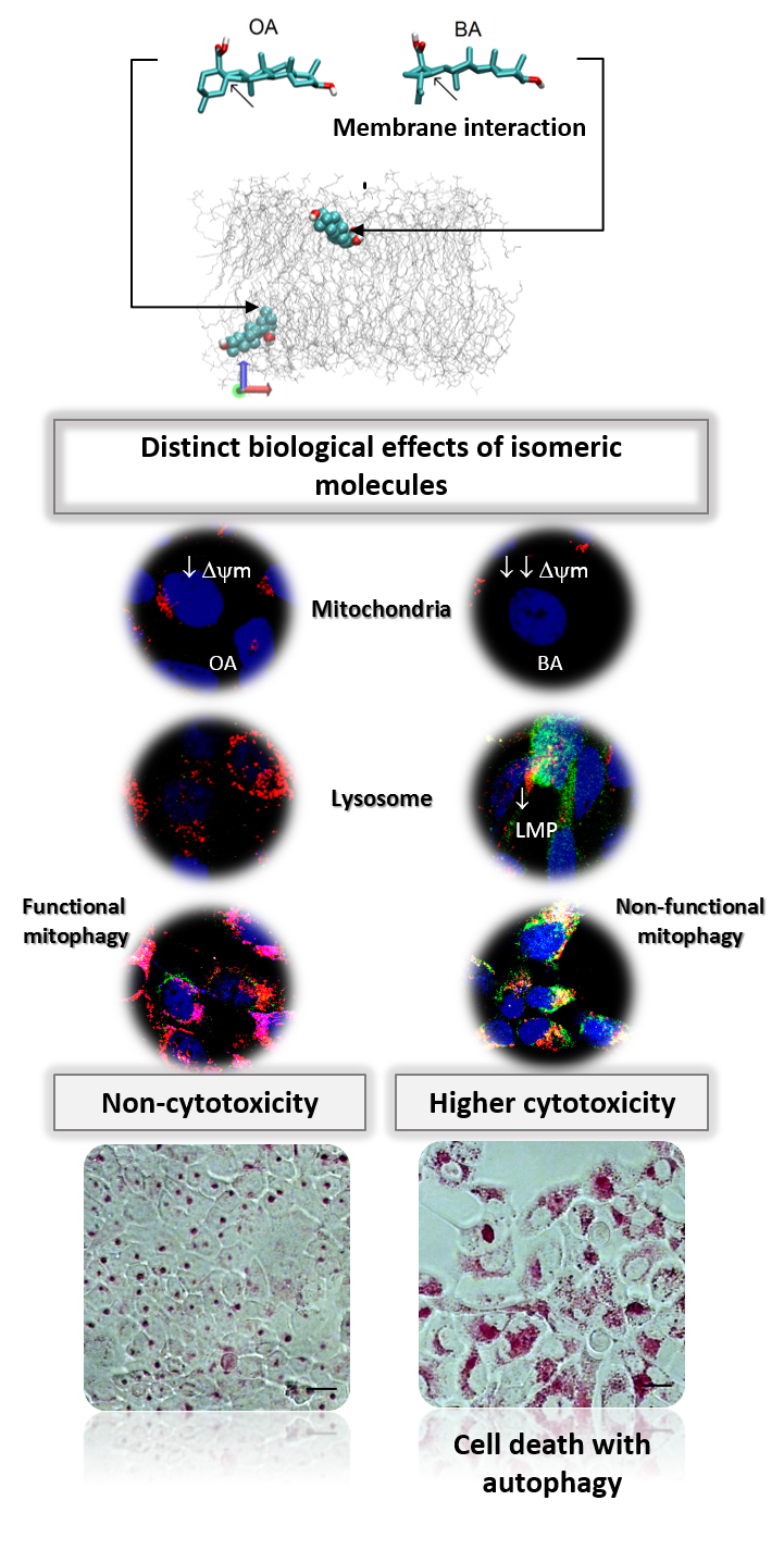
Parallel damage in the mitochondrial and lysosomal membranes leads to cell death with autophagy
An important aim of our CEPID-Redoxoma is to develop diagnostic and therapeutic applications of redox processes. In this context, antioxidant therapies are at the frontline of our interests as a group. In parallel, however, a smaller but nonetheless significant group of strategies aim to explore prooxidant and stress-enhancing effects of distinct interventions, mainly to achieve selective toxicity towards damaged or tumor cells. The group of Prof. Mauricio S. Baptisata, from our CEPID-Redoxoma, has been exploring for more than a decade photo-induced compounds as a means to achieve such type of effects. Interestingly, this group recently provided a significant contribution [1] using triterpenoid pentacyclic molecules which are not photo-active, namely Betulinic acid (BA) and Oleanolic acid (OA). In parallel, these results are helping to understand the cellular responses to several photo-induced processes.

Although being chemical isomers, BA and OA have almost identical physico-chemical properties. However, BA is a lot more cytotoxic. The main difference between these two molecules is their efficiency of membrane interaction. BA binds and damages membranes, an effect that is not observed for OA. This membrane-damaging effect of BA associates with parallel damage on mitochondria and lysosome, turning autophagy into a destructive process. Indeed, the higher cytotoxicity of BA correlated with its stronger efficiency in damaging membrane mimics. OA caused mitochondrial but not lysosomal damage, leading to an autophagy response able to rescue cellular homeostasis. In fact, the response to OA was turned into cell death upon concomitant lysosomal inhibition by chloroquine or bafilomycin-A1. Based on these findings, a new mechanism of cell death was proposed, namely, that autophagy will turn into a destructive outcome when there is parallel damage in mitochondrial and lysosomal membranes.

The transition from the pro-survival to the pro-death roles of autophagy still causes intense debate in the scientific community. These data showed that whether autophagy causes cell rescue or cell death seems to depend on the extent of membrane damage. This concept is likely to provide a basis for the development of new drugs against aggressive cancers. An important outcome of these studies are the ongoing experiments in this group, this time using photoactivable drugs, which are indicating a recurrent scenario of cell death with autophagy upon parallel damage in mitochondria and lysosome.


  1. W. K. Martins, É. T. Costa, M. C. Cruz, B. S. Stolf, R. Miotto, R. M. Cordeiro, M. S. Baptista. Parallel damage in mitochondrial and lysosomal compartments promotes efficient cell death with autophagy: The case of the pentacyclic triterpenoids Scientific Reports, 5: 12425, 2015 | doi: 10.1038/srep12425

Mauricio da Silva Baptista, PhD. Professor at Department of Biochemistry, Institute of Chemistry, University of São Paulo, Brazil

Add new comment

The complex relationship between omega-3 fatty acids and neurodegenerative diseases

Submitted by redoxoma on Sat, 10/31/2015 - 20:10

Redoxoma Highlights by Sayuri Miyamoto

Docosahexaenoic acid (DHA) is an omega-3 fatty acid that is well known by its health-promoting effects. Being highly abundant in the brain, DHA displays essential role in neurological and visual development in infants. In adults, the decline of DHA content in brain has been associated to cognitive impairment and the use of omega-3 supplements have been thought to exert neuroprotective effects. Indeed, some studies indicated that consuming DHA would be beneficial for the prevention of cognitive disorders such as Alzheimer's disease [1]. However, a recent clinical study involving 4000 participants has found no statistically significant difference between the groups that received omega-3 supplements vs the placebo group [2]. Discrepant results on omega-3 benefits has been also reported for another neurodegenerative disease, amyotrophic lateral sclerosis (ALS). While an observational study reported that a diet rich in omega-3 may lower the risk for ALS [3], a contrasting result was observed in another study, in which omega-3 supplementation in the form of eicosapentaenoic acid (EPA) accelerated disease progression in a mouse model of ALS [4]. Animals that were pretreated with EPA showed increased cellular damage and vacuolization within the spinal cord. These effects have been attributed to the presence of increased amounts of lipid-derived oxidation products and superoxide dismutase1 (SOD1) aggregates. To corroborate with the latter hypothesis, a recent study by Appolinario et al. [5], from the group of Sayuri Miyamoto, a principal investigator of CEPID-Redoxoma, demonstrated that DHA induces the aggregation of both wild type and G93A mutant form of apo(without metals)-Cu,Zn-SOD1 in vitro [5]. Interestingly, the study by Appolinario et al. revealed that the oxidized counterpart of DHA (DHA hydroperoxides) induces a distinct pattern of SOD1 aggregation in which aberrant SOD1 dimeric species are produced. Since such SOD1 dimeric aggregates have been thought to be involved in neurodegerative processes, results of this study raise an important question and indicate that further research is needed to understand the role DHA and its oxidized derivatives in ALS disease development. In summary, although epidemiological studies suggest that omega-3 supplements may be beneficial, care should be taken when adopting any supplementation before its efficacy is proven by scientific studies and clinical trials.


  1. M. Fotuhi, P. Mohassel, K. Yaffe. Fish consumption, long-chain omega-3 fatty acids and risk of cognitive decline or Alzheimer disease: a complex association Nature Reviews Neurology, 5, 140-52, 2009 | doi: 10.1038/ncpneuro1044
  2. E. Y. Chew, T. E. Clemons, E. Agrón, L. J. Launer. Effect of omega-3 fatty acids, lutein/zeaxanthin, or other nutrient supplementation on cognitive function The Journal of American Medical Association, 314(8): 791-801, 2015 | doi: 10.1001/jama.2015.9677
  3. K. C. Fitzgerald, É. J. O’Reilly, G. J. Falcone, M. L. McCullough, Y. Park, L. N. Kolonel, A. Ascherio. Dietary ω-3 polyunsaturated fatty acid intake and risk for amyotrophic lateral sclerosis JAMA Neurology, 71(9): 1102-10, 2014 | doi: 10.1001/jamaneurol.2014.1214
  4. P. K. Yip, C. Pizzasegola, S. Gladman, M. L. Biggio, M. Marino, M. Jayasinghe, F. Ullah, S. C. Dyall, A. Malaspina, C. Bendotti, A. Michael-Titus. The omega-3 fatty acid eicosapentaenoic acid accelerates disease progression in a model of amyotrophic lateral sclerosis PLoS One, 8(4): e61626, 2013 | doi: 10.1371/journal.pone.0061626
  5. P. P. Appolinário, D. B. Medinas, A. B. Chaves-Filho, T. Genaro-Mattos, J. R. R. Cussiol, L. E. S. Netto, O. Augusto, S. Miyamoto. Oligomerization of Cu, Zn-superoxide dismutase (SOD1) by docosahexaenoic acid and its hydroperoxides in vitro: aggregation dependence on fatty acid unsaturation and thiols PLoS One, 10(4): e0125146, 2015 | doi: 10.1371/journal.pone.0125146

Sayuri Miyamoto, PhD Associate Professor, Departament of Biochemistry, Institute of Chemistry, University of São Paulo, São Paulo, Brazil.

Add new comment

Together for ever…: Cross-linking of proteins by a ditryptophan bond

Submitted by redoxoma on Sat, 10/31/2015 - 20:09

Redoxoma Highlights by Verônica Paviani

Oxidative modifications of proteins are extensively investigated because proteins are major targets of radicals and oxidants under physiological conditions [1]. The amino acid residues most susceptible to oxidation are the sulfur-containing residues cysteine and methionine and the aromatic residues histidine, phenylalanine, tyrosine and tryptophan. The oxidation of cysteine and methionine residues is reversible and protein-cysteine oxidation is emerging as a fundamental cell regulatory mechanism. In contrast, the oxidation of all other protein residues is irreversible, and may result in loss of protein function, protein aggregation and altered protein turnover, leading to cell and tissue dysfunction.

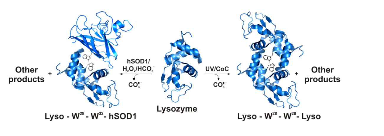
Despite the extensive investigation of the irreversible oxidations undergone by proteins in vitro and in vivo, the products formed from the oxidation of tryptophan residues remain incompletely characterized. However, protein-tryptophan residues have a unique potential to interact with other proteins and cellular structures, and their oxidation may have profound physiological consequences. For instance, our group recently characterized a novel ditryptophan cross-link caused by the recombination of human superoxide dismutase1 (SOD1)-tryptophanyl radicals, which are produced by attack of the carbonate radical (CO3•-) generated during the enzyme´s bicarbonate-dependent peroxidase activity [2]. In addition, it was demonstrated that this cross-link contributes to triggering the non-amyloid aggregation of hSOD1, a process that may be involved in the pathogenic mechanism of the nuerodegenerative disease amyotrophic lateral sclerosis [3]. Now, we show that attack of the CO3•- on lysozyme produces lysozyme and lysozyme-hSOD dimers cross-linked by a ditryptophan bond. We also show that the same lysozyme dimer is produced by UV irradiation of the enzyme [4]. In addition to confirming that the CO3•- tends to promote protein cross-links and aggregation, the results show that UV light can act similarly. Since the produced protein lesions are resistant to cellular repair, they are likely to play a role in the pathogenic mechanism of protein aggregation diseases.

Cross-linking of proteins by a ditryptophan bond


  1. M. J. Davies. The oxidative environment and protein damage Biochimica et Biophysica Acta, 170: 93-109, 2005 | doi: 10.1016/j.bbapap.2004.08.007
  2. D. B. Medinas, F. C. Gozzo, L. F. A. Santos, A. H. Iglesias, O. Augusto. A ditryptophan cross-link is responsible for the covalent dimerization of human superoxide dismutase 1 during its bicarbonate-dependent peroxidase activity Free Radical Biology & Medicine, 49: 1046-53, 2010 | doi: 10.1016/j.freeradbiomed.2010.06.018
  3. F. R. Coelho, A. Iqbal, E. Linares, D. F. Silva, F. S. Lima, I. M. Cuccovia, O. Augusto. Oxidation of the tryptophan 32 residue of human superoxide dismutase1 caused by Its bicarbonate-dependent peroxidase activity triggers the non-amyloid aggregation of the enzyme The Journal of Biological Chemistry, 289: 30690-701, 2014 | doi: 10.1016/j.bbapap.2004.08.007
  4. V. Paviani, R. F. Queiroz, E. F. Marques, P. Di Mascio, O. Augusto. Production of lysozyme and lysozyme-superoxide dismutase dimers bound by a ditryptophan cross-link in carbonate radical-treated lysozyme Free Radical Biology & Medicine, in press. | doi: 10.1016/j.freeradbiomed.2015.07.015

Verônica Paviani, MS Departament of Biochemistry, Institute of Chemistry, University of São Paulo, São Paulo, Brazil.

Add new comment